

Rat Bone Marrow Dendritic Cells
Cat.No.: CSC-C4973J
Species: Rat
Source: Bone Marrow
Cell Type: Dendritic Cell
- Specification
- Background
- Scientific Data
- Q & A
- Customer Review
Never can cryopreserved cells be kept at -20 °C.
Rat bone marrow dendritic cells are the main antigen presenting cells obtained from rat bone marrow progenitor cells in well specified culture conditions. They exhibit a typical dendritic shape and express surface markers related to antigen presentation and T-cell activation, making them a commonly utilized model to study innate and adaptive immune responses. Dendritic cells are professional antigen-presenting cells that play a primary role in the initiation and regulation of immune responses, including the capture, processing and presentation of antigens to T lymphocytes. These cells are often utilized to examine dendritic cell maturation, antigen uptake and presentation, cytokine generation and interactions with T cells and other immune cell types. These cells may influence downstream immunological activation through upregulation of co-stimulatory molecules and secretion of immunoregulatory cytokines in response to stimulation.
Rat bone marrow dendritic cells are extensively applied in immunological control, transplantation immunology, viral illnesses, vaccine development and inflammatory diseases. They may also serve as a model for experimental evaluation of immunomodulatory drugs, biomaterials and cell-based treatment strategies. Furthermore, these cells are commonly used in mixed lymphocyte reaction assays and antigen-specific T-cell activation investigations to examine processes of immunological tolerance and immune stimulation.
Lactobacillus johnsonii N6.2 Polarizes Bone Marrow Dendritic Cells Toward an Immature-Migratory Phenotype
Gut dysbiosis has been related to systemic inflammatory illnesses, however the mechanisms are not yet known. Phospholipids (PLs) are precursor of inflammatory and resolving mediators and their dysregulation is linked to chronic diseases. Researchers purified lipids from L. johnsonii N6.2 using bioassay-guided fractionation to clarify how gut bacteria lipids alter immunity. Cuaycal et al. stimulated rat bone marrow dendritic cells (BMDCs) with total lipids and PL fractions, and subsequently performed RNA sequencing to find transcriptional markers.
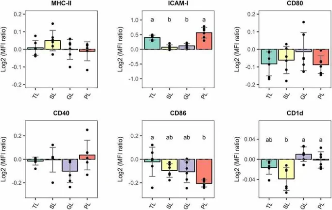
DC maturation is linked to elevated surface expression of MHC, costimulatory molecules and migratory markers facilitating effective T-cell priming. To evaluate the immunomodulatory effects of Lactobacillus johnsoniiN6.2, BMDCs were treated with total lipids (TLs) or lipid fractions (sterolipids [SL], glycolipids [GL], phospholipids [PL]). Flow cytometry quantification of surface marker expression shown as log 2 fold change relative to vehicle control (Fig. 1). PLs stimulation significantly decreased CD86 expression (p<0.05) but did not change MHC-II, CD80 or CD40 level. Instead, the migratory marker ICAM-1 was considerably elevated (p< 0.05) by both TLs and PLs. Moreover, SL stimulated cells showed considerably lower levels of the lipid presenting molecule CD1d compared to GL and PL stimulated BMDCs.
After 6 hours of stimulation, the overall effect of L. johnsoniiN6.2 PLs on BMDCs was an immature-migratory phenotype with unchanged surface levels of MHC-II and costimulatory markers (CD80, CD86, CD40) and upregulation of the migratory marker ICAM-1. This shows that certain bacterial lipid fractions stimulate a tolerogenic-like DC profile that is transcriptionally primed for migration rather than strong activation.
Ask a Question
Write your own review
- You May Also Need
Description: The thoracic aorta is located in the chest cavity and gives off arteries that branch to the esophagus, pericardium, lungs, and trachea. The thoracic aorta can be subdivided into the ascending aorta, ...
Description: Rat Podocytes are isolated from normal rat kidney. The cells are characterized by immunofluorescence with antibodies specific to podocin, Ang1, Nephrin, ACTN4, NPHS2. T25 flasks is required for cell ...
Description: Rat Bronchial Smooth Muscle Cells are isolated from normal rat bronchi tissue. Rat Bronchial Smooth Muscle Cells are characterized by immunofluorescence with antibodies specific to alpha-actin. T25 ...
Description: Guinea Pig Endothelial Cells from Creative Bioarray are isolated from guinea pig tissue. Prior to shipping, cells at passage 2 are detached from flasks and immediately cryopreserved in vials. Each ...
Description: Rat Lung Epithelial Cells are isolated from normal rat lung tissue. The cells are characterized by immunofluorescence with antibodies specific to CK-18, CK-19. T25 flasks is required for cell ...
Description: Rat Vein Endothelial Cells from Creative Bioarray are isolated from inferior vena cava tissue of 6-8 week old laboratory Sprague-Dawley rat. Rat Vein Endothelial Cells are grown in T75 tissue culture ...