
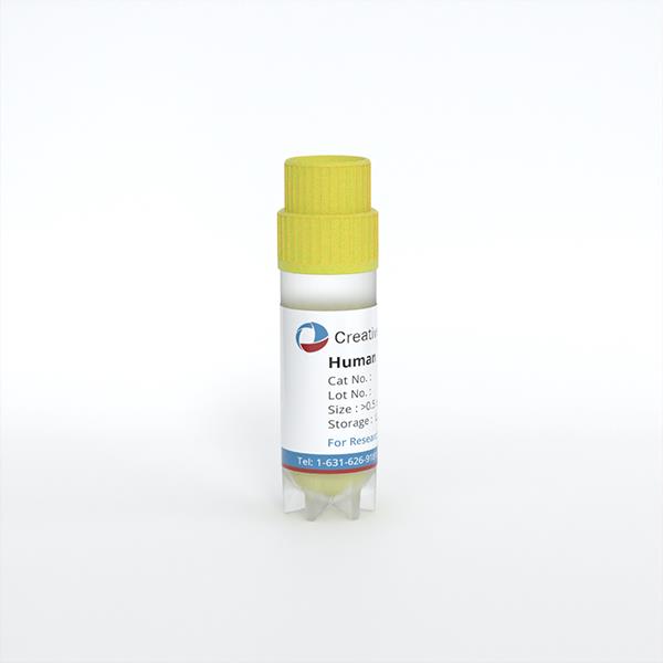
Human Spleen Microvascular Endothelial Cells
Cat.No.: CSC-C4868L
Species: Human
Source: Spleen
Cell Type: Endothelial Cell; Microvascular Cell
- Specification
- Background
- Scientific Data
- Q & A
- Customer Review
Never can cryopreserved cells be kept at -20 °C.
Human Spleen Microvascular Endothelial Cells are primary endothelial cells obtained from the microvasculature of human spleen tissue. These cells line tiny blood arteries in the spleen and help to maintain vascular integrity, regulate permeability, and facilitate the interchange of cells and chemicals between the circulation and splenic tissue. In culture, they usually have a cobblestone-like shape and express endothelial markers including CD31 (PECAM-1), von Willebrand factor (vWF), and VE-cadherin, which are related with endothelial structure and function.
Splenic microvascular endothelial cells regulate the immune system by interacting with circulating leukocytes and facilitating their adhesion and motility. Given the spleen's function in immunological surveillance and blood filtration, these cells are frequently utilized to investigate leukocyte trafficking, inflammatory signaling, and endothelial responses to cytokines or infections. They may also be used in investigations on vascular dysfunction or immune-related illnesses.
CD14dim/CD16++ Monocytes Induce VWF:Ag Release from Splenic ECs, in vitro
Desmopressin (DDAVP) treats bleeding disorders by releasing von Willebrand factor (VWF), but its mechanism is unclear as endothelial AVPR2 receptors remain undemonstrated. The spleen, a major FVIII-producing organ with distinct monocyte reservoirs, may mediate this effect. Salarilak et al. examined a splenectomised haemophilia patient with impaired DDAVP response, then used in vitro co-culture experiments to assess VWF release from DDAVP-treated human splenic endothelial cells (HSECs) with different monocyte subtypes.
Neonatal and adult HSECs co-produced FVIII and VWF (~0.2 mU/cm² FVIII:C and 3 mU/cm² VWF:Ag in 48-h supernatants up to passage 4), declining thereafter until FVIII:C was undetectable at passage 6. Confluent HSECs in monoculture or co-culture with PBMCs, classical (CD14/CD16-), or non-classical (CD14dim/CD16) monocytes were treated with 100 ng/ml DDAVP for 1 h. DDAVP did not induce VWF or P-selectin release compared to untreated cells, while PMA significantly induced VWF:Ag release. Proximity of PBMCs or classical monocytes did not influence this. However, direct exposure of CD14dim/CD16++ monocytes to HSECs enabled DDAVP to significantly increase VWF:Ag in supernatants.
As platelet-activating factor (PAF) was proposed as a potential signal transmitter from CD14dim/CD16++ monocytes, HSECs were treated with pure PAF (12 ng/ml-40 μg/ml). Neither PAF nor DDAVP induced VWF:Ag release, while PMA remained effective, suggesting PAF alone does not mediate the monocyte-driven DDAVP effect.
Ask a Question
Write your own review
Description: HSF from Creative Bioarray are isolated from human spleen tissue. HSF are cryopreserved at primary culture and delivered frozen. Each vial contains >5 x 10^5 cells in 1 ml volume. HSF are ...
Description: Human Spleen Epithelial Cells are isolated from normal human spleen tissue.
Description: Human Spleen Fibroblasts are isolated from normal human spleen tissue.
Description: HSEC from Creative Bioarray are isolated from human spleen. HSEC are cryopreserved at passage one and delivered frozen. Each vial contains >5 x 10⁵ cells in 1 ml volume. HSEC are characterized by ...
Description: Human splenocytes from Creative Bioarray are isolated from human spleen tissues. Each vial contains 5x10^6 cells per ml and is delivered frozen. Human splenocytes are negative for bacteria, yeast, ...