
Human Spleen Fibroblasts
Cat.No.: CSC-C4853L
Species: Human
Source: Spleen
Cell Type: Fibroblast
- Specification
- Background
- Scientific Data
- Q & A
- Customer Review
Never can cryopreserved cells be kept at -20 °C.
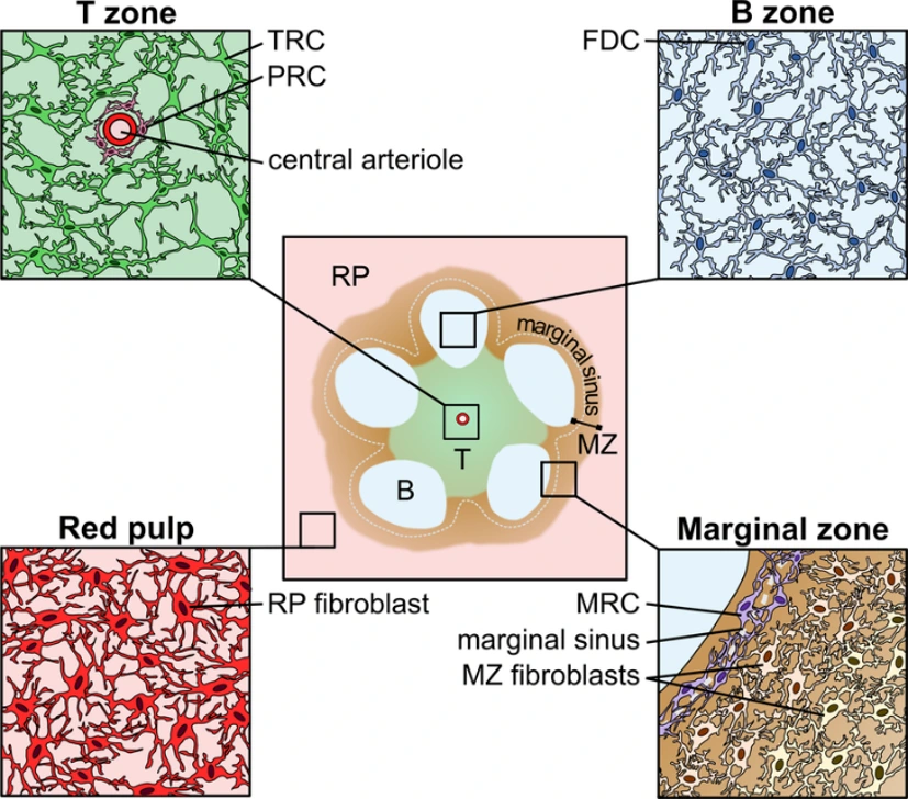
Human Spleen Fibroblasts are primary stromal cells derived from human spleen tissue. They constitute a structurally and functionally unique fibroblast subset that has been molded by the organ's specialized function in blood filtration and immune system regulation. In contrast to fibroblasts from many other tissues, those found in the spleen operate in a milieu continuously exposed to circulating immune cells and antigens, conferring upon them robust immunomodulatory and matrix-organizing functions.
Human spleen fibroblasts share the spindle-shaped morphology characteristic of fibroblasts but are particularly adept at producing and remodeling extracellular matrix components to support the splenic red and white pulp compartments. In the spleen, these cells closely interact with lymphocytes, macrophages, and endothelial cells to organize immune cell niches and guide cell migration. They are highly sensitive to inflammatory cytokines and immune cell-derived cues, rapidly modulating their behavior in response to immune activation or systemic inflammation. These cells have been extensively utilized to study stromal-immune cell interactions, immune tissue remodeling, and inflammation-induced fibrosis. Due to their immune-aware phenotype, these cells are particularly useful for investigating splenic function, immune disorders, hematologic diseases, and for testing therapies that target stromal regulation in immune organs.
The Impact of Photolithographic Resin Products on HSF Mammalian Cell Culture
Composite materials based on PMMA and carbon nanoparticles are widely used in various fields. While carbon nanotubes and nano-dots are commonly used, they are expensive and difficult to obtain. Gudkov et al. explored the use of easily obtainable and low-cost amorphous carbon nanoparticles to modify PMMA, investigating their physico-chemical and biological characteristics.
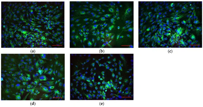
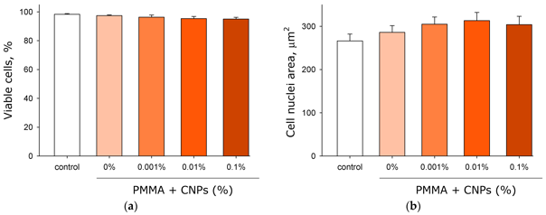
The impact of photolithographic resin products on HSF mammalian cell culture was studied (Fig. 1). Microscopic analysis showed no significant differences between cell groups. Cells adhered and spread equally well on both culture glass and fabricated samples with or without CNPs. Cell viability was assessed using fluorescence microscopy (Fig. 2a). Most cells appeared viable, with over 95% viability in all groups, and no significant differences between groups. The effect of the fabricated samples on nuclear size was also examined (Fig. 2b). Nuclear size did not differ significantly among the experimental groups.
Ask a Question
Write your own review

Description: HSF from Creative Bioarray are isolated from human spleen tissue. HSF are cryopreserved at primary culture and delivered frozen. Each vial contains >5 x 10^5 cells in 1 ml volume. HSF are ...
Description: Human Spleen Epithelial Cells are isolated from normal human spleen tissue.
Description: HSEC from Creative Bioarray are isolated from human spleen. HSEC are cryopreserved at passage one and delivered frozen. Each vial contains >5 x 10⁵ cells in 1 ml volume. HSEC are characterized by ...
Description: Human splenocytes from Creative Bioarray are isolated from human spleen tissues. Each vial contains 5x10^6 cells per ml and is delivered frozen. Human splenocytes are negative for bacteria, yeast, ...
Description: Human Spleen Microvascular Endothelial Cells are isolated from human spleen tissue.