
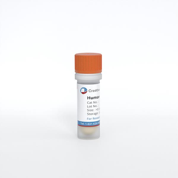
Human Lung Adenocarcinoma Cancer Associated Fibroblasts
Cat.No.: CSC-C30035J
Species: Human
Source: Lung
Cell Type: Fibroblast
- Specification
- Background
- Scientific Data
- Q & A
- Customer Review
Human Lung Adenocarcinoma Cancer-Associated Fibroblasts (hLUAD-CAFs) are primary stromal cells isolated directly from patient tumor specimens, representing the most abundant and functionally dominant non-malignant constituents of the lung adenocarcinoma tumor microenvironment (TME). Unlike immortalized fibroblast lines or animal xenografts, hLUAD-CAFs preserve patient-specific genetic and epigenetic landscapes, sustain native activated myofibroblast phenotypes, and authentically recapitulate the intricate paracrine signaling networks that govern tumor progression, extracellular matrix (ECM) desmoplasia, and therapeutic resistance.
These cells constitutively express established CAF markers—including α-SMA, FAP-α, vimentin, and PDGFR-β—while secreting a potent array of tumor-modulating mediators such as TGF-β1, IL-6, CXCL12/SDF-1, and matrix metalloproteinases. Through dynamic ECM remodeling and bidirectional crosstalk with malignant and immune compartments, hLUAD-CAFs actively promote cancer cell proliferation, angiogenesis, epithelial-mesenchymal transition, immunosuppressive macrophage polarization, and regulatory T-cell recruitment. Contemporary single-cell transcriptomic analyses have further unveiled substantial inter-patient heterogeneity among hLUAD-CAF subpopulations, including myofibroblastic and inflammatory subsets, underscoring their indispensable value in personalized biomarker discovery, patient stratification, and subtype-specific therapeutic targeting.
As a physiologically relevant ex vivo platform, hLUAD-CAFs offer distinct and unparalleled experimental advantages: native secretory fidelity, superior translational predictability across diverse in vitro systems, and seamless compatibility with co-culture platforms, 3D organoid models, and high-throughput drug screening workflows. Moreover, their relative genetic stability compared to malignant cells renders them robust, druggable targets for mechanism-driven validation and stroma-directed therapeutic discovery.
Cancer-Associated Fibroblast-Derived SOD3 Enhances Lymphangiogenesis to Drive Metastasis in Lung Adenocarcinoma
Despite advancements in diagnostic and therapeutic strategies, lung adenocarcinoma (LUAD) remains a leading cause of cancer-related mortality due to its aggressive metastatic potential. Extracellular superoxide dismutase (SOD3) is an antioxidant enzyme that regulates oxidative stress and is regarded as a tumor suppressor. However, its molecular mechanisms within the tumor microenvironment are poorly understood. In this study, Oo, May Wathone, et al. report a breakthrough in uncovering the role of SOD3 derived from cancer-associated fibroblasts (CAFs) in LUAD.
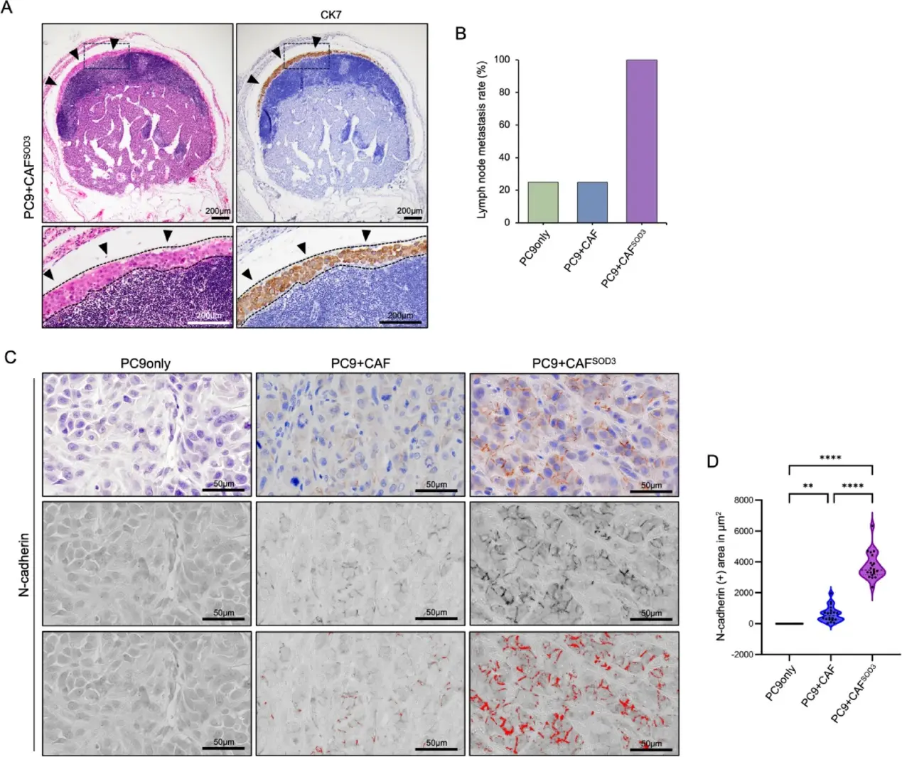
Using a mouse xenograft LUAD model transplanted with human-derived CAF, they showed that overexpression of SOD3 in CAF induces lymphangiogenesis, resulting in tumor metastasis in the lymph nodes. Mechanistically, transcriptome analysis revealed that SOD3 overexpression in CAF enhanced cancer exacerbation-related genes, particularly angiogenesis, lymph vessel development, collagen degradation, and epithelial-mesenchymal transition. These findings suggest that SOD3 may be a prognostic factor for LUAD patients, and SOD3-expressing CAF could be a potential therapeutic target to disrupt the stroma-tumor alliance relation in tumor development and progression.
Ask a Question
Write your own review
Description: Human Pulmonary Artery Fibroblasts are isolated from normal human pulmonary artery.
Description: Primary Human Fetal Lung Fibroblast Cells were initiated from human lung tissue (normal 25 week gestation).These cells were originated using Complete Serum-Free Medium Kit With SuperFuel™, are ...
Description: GFP-Human Lung Airway Smooth Muscle Cells (GFP-HLAWSMCs) provided by Creative Bioarray are puromycin-selected from primary human lung airway smooth muscle cells infected with lentivirus expressing ...
Description: ACE2-GFP Expressing Human Pulmonary Artery Endothelial Cells (ACE2-GFP-HPAECs) provided by Creative Bioarray are puromycin-selected from Human Pulmonary Artery Endothelial Cells infected with ...
Description: HTSMC from Creative Bioarray Research are isolated from human trachea. HTSMC are cryopreserved at secondary culture and delivered frozen. Each vial contains >5 x 10^5 cells in 1 ml volume. HTSMC are ...
Description: The human type 1 alveolar epithelial cells from Creative Bioarray are isolated from human alveolar tissue. These cells are shipped at passage 2 and maintained in SuperCult® human type 1 alveolar ...