Canine Lung Fibroblasts
Cat.No.: CSC-C4810L
Species: Dog
Source: Lung
Cell Type: Fibroblast
- Specification
- Background
- Scientific Data
- Q & A
- Customer Review
Never can cryopreserved cells be kept at -20 °C.
Canine Lung Fibroblasts are primary cells isolated from the pulmonary parenchyma of healthy dogs, most commonly sourced from the lung lobes of adult or young donors. These cells exhibit the classic spindle-shaped, fibroblast-like morphology and grow as adherent monolayers. Their primary physiological role involves synthesizing and remodeling the extracellular matrix (ECM) of the lung interstitium, maintaining structural integrity and facilitating tissue repair.
In vitro, they are maintained in a nutrient-rich basal medium supplemented with serum and essential amino acids under standard conditions (37°C, 5% CO₂). To preserve their phenotype and prevent spontaneous differentiation, they are typically passaged via trypsinization at 70-80% confluence and are best utilized within early passages (P3-P8).
These fibroblasts are a cornerstone model in veterinary respiratory research, particularly for studying pulmonary fibrosis (PF), where they are activated to investigate the mechanisms of fibroblast-to-myofibroblast transition (FMT) and excessive collagen deposition. They are also extensively used to model Acute Respiratory Distress Syndrome (ARDS) and Inflammatory Lung Injury, examining how fibroblasts respond to pro-inflammatory cytokines (e.g., IL-1β, TGF-β) and environmental toxins. Furthermore, Canine Lung Fibroblasts serve as a vital preclinical platform for evaluating the cytotoxicity and safety profiles of inhaled drugs, nebulized therapies, and medical devices intended for veterinary and translational human applications.
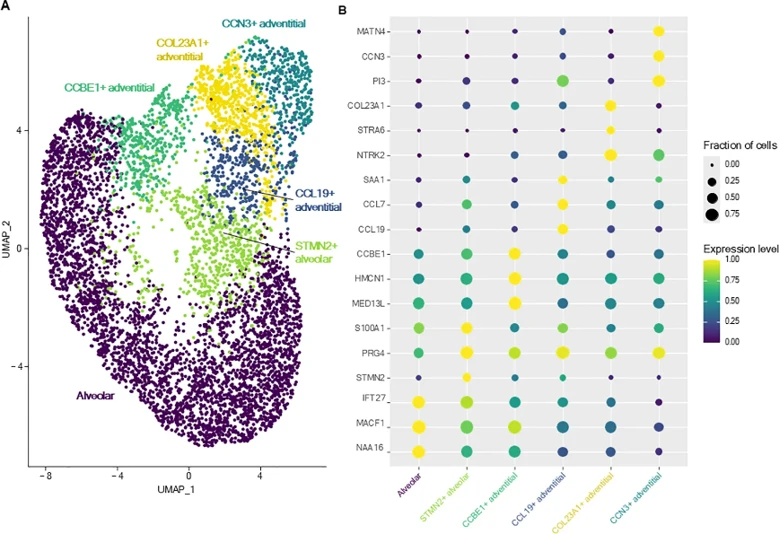
A Single-Cell RNA Sequencing Atlas of the Healthy Canine Lung
While single-cell RNA sequencing has profoundly advanced our understanding of human lung biology, the cellular architecture of the healthy canine lung remains largely undefined despite the translational importance of dogs in modeling human respiratory disease. To address this, Rizzoli et al. constructed the first comprehensive single-cell atlas of the healthy canine lung, focusing specifically on the fibroblast landscape.
Sub-clustering analysis identified six transcriptionally distinct fibroblast populations, which were categorized into alveolar (distal) and adventitial (proximal) subtypes based on established mesenchymal classifications (Fig. 1). Alveolar fibroblasts were characterized by canonical markers such as COL13A1, WNT2, and NPNT. Notably, they identified a unique subset, termed STMN2+ alveolar fibroblasts, which overexpressed STMN2, PRG4, and IL33, alongside cytokine and chemokine genes like CCL19 and CXCL12. Gene Ontology analysis revealed that this subset was significantly enriched for inflammatory response and cytokine-mediated signaling pathways. Adventitial fibroblasts, defined by COL14A1, GLI1, and DCN expression, were further resolved into four specialized clusters. These included CCL19+ fibroblasts, which exhibited pro-inflammatory profiles; CCN3+ fibroblasts, associated with cell migration and proliferation; COL23A1+ fibroblasts, implicated in organ morphogenesis; and CCBE1+ fibroblasts, linked to circulatory system development. Together, these findings demonstrate that canine lung fibroblasts possess profound transcriptional and functional heterogeneity, providing a critical foundation for future comparative studies in pulmonary disease.
Ask a Question
Write your own review
Description: Dog Liver Endothelial Cells from Creative Bioarray are isolated from tissue of dog liver. Dog Liver Endothelial Cells are grown in T25 tissue culture flasks pre-coated with gelatin-based coating ...

Description: Canine Astrocytes from Creative Bioarray are isolated from canine brain tissue. The method we use to isolate canine astrocytes were developed based on a combination of established and our proprietary ...
Description: Canine Mammary Microvascular Endothelial Cells from Creative Bioarray are isolated from breast of pathogen-free laboratory Canine. Canine Mammary Microvascular Endothelial Cells are grown in T25 ...
Description: Canine Chondrocytes (CnC) provided by Creative Bioarray are isolated from normal canine articular cartilage tissue. The cells are frozen at passage 1 and each vial contains at least 0.5*10^6 cells. ...
Description: Canine Pancreatic Microvascular Endothelial Cells from Creative Bioarray are isolated from Pancreatic Microvascular of pathogen-free laboratory Canine. Canine Pancreatic Microvascular Endothelial ...
Description: Canine Prostate Microvascular Endothelial Cells from Creative Bioarray are isolated from prostate of pathogen-free laboratory Canine. Canine Prostate Microvascular Endothelial Cells are grown in T25 ...