
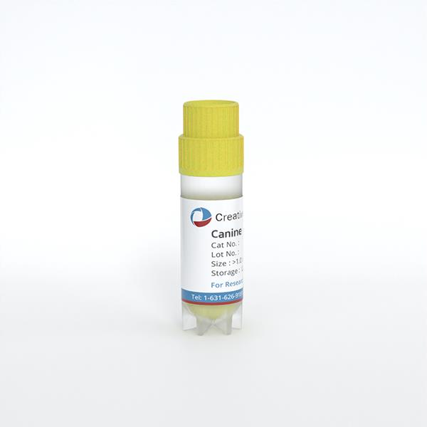
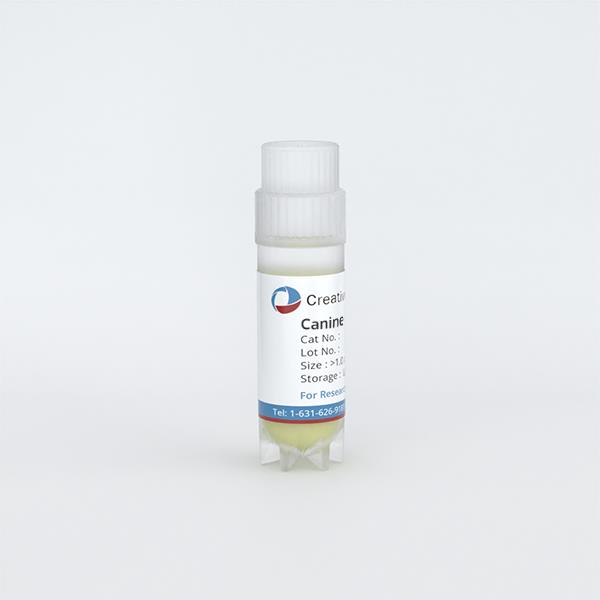
Canine Dermal Fibroblasts-Neonatal
Cat.No.: CSC-C4806L
Species: Dog
Source: Dermis; Skin
Cell Type: Fibroblast
- Specification
- Background
- Scientific Data
- Q & A
- Customer Review
Never can cryopreserved cells be kept at -20 °C.
Canine Dermal Fibroblasts from neonatal donors are primary cells extracted from the dermis of newborn puppy skin (usually <7 days old). They are spindle-shaped, fibroblast-like cells which develop as an adherent monolayer and synthesize the dermal extracellular matrix, mainly type I and III collagen, elastin and fibronectin, making them vital for wound healing and tissue remodeling.
The neonatal canine fibroblasts demonstrate increased attachment and a stronger proliferative capability compared to adult fibroblasts in culture and are cultured under standard conditions (37°C, 5% CO2) in a rich basal medium (e.g., DMEM or MEM) containing serum and L-glutamine. They are usually passaged at 70-80% confluency with trypsin/EDTA and advised to be used in early passages (P2-P6) to maintain phenotypic and to avoid replicative senescence.
These cells are commonly employed in the investigation of veterinary dermatology and wound repair, including burn healing, scar formation and fibroblast-keratinocyte cross-talk studies. They also represent a biologically relevant substrate for cutaneous toxicity screening of topical formulations, cytotoxicity assays of veterinary pharmaceuticals, and as a control normal counterpart in comparative studies with canine cutaneous tumor cells (e.g. mast cell tumors, soft tissue sarcomas). In addition, newborn canine fibroblasts are used in stem cell and tissue engineering applications, for example, to seed decellularized dermal scaffolds for regenerative medicine in veterinary and translational animal models.
Chitosan Coating Mitigates SPION Cytotoxicity but Promotes Aggregation in Biological Media
Superparamagnetic iron oxide nanoparticles (SPIONs) hold promise for biomedical applications, but balancing colloidal stability, biocompatibility, and magnetic performance remains challenging. Mistral et al. investigated how chitosan (CS) coatings with varying degrees of acetylation (DA) modulate SPION behavior in biological systems.
Optical microscopy revealed that SPIONs aggregated in cell culture medium, with aggregation intensity correlating positively with DA and coating mass fraction. Aggregates preferentially localized to cellular layers rather than cell-free spaces, appearing to interact with or be internalized by canine fibroblasts. Contrary to expectations, CS acted as an adhesive rather than a steric stabilizer, promoting heterogeneous particle distribution.
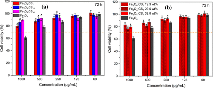
Cytocompatibility assessed via CCK-8 assays showed that cell viability increased with SPION dilution (Fig. 1). At concentrations ≤125 µg/mL, all formulations maintained >80% viability across 24-72 hours. Uncoated magnetite SPIONs exhibited significant toxicity at 1 mg/mL (~60% viability), whereas CS coating mitigated this effect regardless of DA or coating thickness. However, these results must be interpreted cautiously, as SPION aggregation creates spatial heterogeneity: aggregates may resist internalization or create artificial cell-particle contact zones, potentially biasing viability assessments. Future work should focus on achieving stable dispersions at high coating densities to decouple aggregation effects from intrinsic cytocompatibility.
Ask a Question
Write your own review
Description: Dog Liver Endothelial Cells from Creative Bioarray are isolated from tissue of dog liver. Dog Liver Endothelial Cells are grown in T25 tissue culture flasks pre-coated with gelatin-based coating ...
Description: Canine Astrocytes from Creative Bioarray are isolated from canine brain tissue. The method we use to isolate canine astrocytes were developed based on a combination of established and our proprietary ...
Description: Canine Mammary Microvascular Endothelial Cells from Creative Bioarray are isolated from breast of pathogen-free laboratory Canine. Canine Mammary Microvascular Endothelial Cells are grown in T25 ...
Description: Canine Chondrocytes (CnC) provided by Creative Bioarray are isolated from normal canine articular cartilage tissue. The cells are frozen at passage 1 and each vial contains at least 0.5*10^6 cells. ...
Description: Canine Pancreatic Microvascular Endothelial Cells from Creative Bioarray are isolated from Pancreatic Microvascular of pathogen-free laboratory Canine. Canine Pancreatic Microvascular Endothelial ...
Description: Canine Prostate Microvascular Endothelial Cells from Creative Bioarray are isolated from prostate of pathogen-free laboratory Canine. Canine Prostate Microvascular Endothelial Cells are grown in T25 ...