

Rat Primary Dermal Fibroblasts-adult
Cat.No.: CSC-C4169X
Species: Rat
Source: Dermis; Skin
Cell Type: Fibroblast
- Specification
- Background
- Scientific Data
- Q & A
- Customer Review
Cells are negative for bacteria, yeast, fungi, and mycoplasma. Rat Primary Dermal Fibroblasts are tested for expression of marker using the antibody of anti-FSP1/S100A4 by immunofluorescence staining and can be expanded by 2-4 passages at a split ratio of 1:2 under the cell culture conditions specified by Creative Bioarray. Repeated freezing and thawing of cells is not recommended.
Standard biochemical procedures performed with fibroblast cultures include the assays of cell to cell interaction, PCR, Western blotting, immunoprecipitation, immunofluorescent staining, immunofluorescent flow cytometry or generating cell derivatives for desired research applications.
Rat primary dermal fibroblasts are non-immortalized mesenchymal cells isolated from the dermis of adult rats. They retain a normal diploid karyotype, spindle-shaped morphology, and robust expression of vimentin, faithfully recapitulating in vivo fibroblast biology without the aberrant signaling of transformed lines. Their adult origin confers intrinsic age-related characteristics, including defined proliferative capacity, susceptibility to replicative senescence, and a mature extracellular matrix (ECM) secretory profile, making them a superior model for studying chronological and photoaging.
A defining advantage is their potent responsiveness to TGF-β1, which drives efficient transdifferentiation into α-smooth muscle actin-positive myofibroblasts, providing a physiologically authentic system for dissecting fibrosis, scar formation, and wound contraction. These cells constitutively secrete abundant type I and III collagen, fibronectin, and matrix metalloproteinases, enabling the assembly of three-dimensional endogenous ECM for mechanotransduction and tissue remodeling studies. They are amenable to lentiviral transduction, siRNA-mediated gene silencing, and co-culture with keratinocytes or macrophages to model dermal-epidermal crosstalk and inflammation. Importantly, they retain functional mechanosensing pathways, allowing investigation of stiffness-dependent fibroblast activation.
Collectively, their physiological fidelity, myofibroblast differentiation capacity, and experimental tractability make them an indispensable platform for exploring dermal matrix biology, fibrotic disease mechanisms, and anti-fibrotic drug screening.
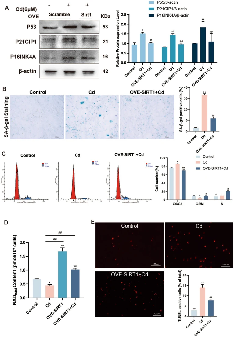
SIRT1 Regulates Dermal Fibroblast Senescence Induced by Cadmium Exposure
Skin aging is a complex, multifactorial biological process that can be significantly accelerated by environmental toxicants such as cadmium (Cd), a highly toxic and ubiquitous heavy metal. Although the broad cytotoxic impacts of Cd have been extensively reported, a comprehensive understanding of the precise molecular pathways underlying Cd-induced skin senescence is still lacking. In this study, Zhou, Dehui, et al. investigated the protective role of Sirtuin 1 (SIRT1), a highly conserved nicotinamide adenine dinucleotide (NAD+)-dependent deacetylase that functions as a master regulator of mitochondrial homeostasis and cellular defense mechanisms.
An in vitro model was established using primary rat dermal fibroblasts and C3H/10 T1/2 cells, with SIRT1 levels modulated via lentiviral-mediated overexpression. The results demonstrate that Cd exposure elevates reactive oxygen species (ROS), disrupts mitochondrial integrity, and activates DNA damage responses, collectively driving cellular senescence. SIRT1 was shown to exert protective effects through the deacetylation of key substrates such as P53 and SOD2, thereby restoring redox balance and promoting DNA repair. The elevation of SIRT1 expression markedly mitigated mitochondrial impairments, senescent phenotypes, and apoptotic features triggered by Cd exposure.
Ask a Question
Write your own review
Description: The thoracic aorta is located in the chest cavity and gives off arteries that branch to the esophagus, pericardium, lungs, and trachea. The thoracic aorta can be subdivided into the ascending aorta, ...
Description: Rat Podocytes are isolated from normal rat kidney. The cells are characterized by immunofluorescence with antibodies specific to podocin, Ang1, Nephrin, ACTN4, NPHS2. T25 flasks is required for cell ...
Description: Rat Bronchial Smooth Muscle Cells are isolated from normal rat bronchi tissue. Rat Bronchial Smooth Muscle Cells are characterized by immunofluorescence with antibodies specific to alpha-actin. T25 ...
Description: Guinea Pig Endothelial Cells from Creative Bioarray are isolated from guinea pig tissue. Prior to shipping, cells at passage 2 are detached from flasks and immediately cryopreserved in vials. Each ...
Description: Rat Lung Epithelial Cells are isolated from normal rat lung tissue. The cells are characterized by immunofluorescence with antibodies specific to CK-18, CK-19. T25 flasks is required for cell ...
Description: Rat Vein Endothelial Cells from Creative Bioarray are isolated from inferior vena cava tissue of 6-8 week old laboratory Sprague-Dawley rat. Rat Vein Endothelial Cells are grown in T75 tissue culture ...