
Rat Dorsal Root Ganglion Neurons
Cat.No.: CSC-C8061L
Species: Rat
Source: Spinal Cord
Cell Type: Neuron
- Specification
- Background
- Scientific Data
- Q & A
- Customer Review
A ganglion is a group of nerve cells forming a nerve center, especially one located outside the brain or spinal cord. Dorsal root ganglion, also called spinal ganglion, is the ganglion of the posterior root of each spinal segmental nerve, containing the cell bodies of the unipolar primary sensory neurons. Dorsal root ganglion cells are pseudounipolar cells. Pseudounipolar cells have 2 axons rather than an axon and dendrite. One axon extends centrally toward the spinal cord; the other axon extends toward the skin or muscle.
Applications:
Receptor signaling studies
Gene ex
Intracellular transport studies
Electrophysiology
Metabolic pathway studies
Neurotoxicity
Drug screening
Disease studies
Rat dorsal root ganglion (DRG) neurons are the primary sensory afferents responsible for transducing diverse somatosensory stimuli-including noxious, thermal, mechanical, and proprioceptive signals-from peripheral tissues to the spinal cord. Cultured embryonic or adult rat DRG neurons faithfully preserve native electrophysiological signatures, receptor repertoires, and neurochemical identities, providing a physiologically authentic system that markedly surpasses immortalized neuronal lines lacking full ion-channel expression.
A cardinal advantage is the intrinsic phenotypic heterogeneity retained in culture: small-diameter neurons co-express nociceptive markers such as TRPV1, Nav1.8, CGRP, and substance P, while large-diameter, neurofilament-rich neurons model mechanotransduction and proprioception, enabling cell-type-specific dissection of sensory modalities within a single preparation. Their spherical somata are ideally suited for high-resolution patch-clamp recording of tetrodotoxin-resistant sodium currents, voltage-gated calcium channels, and ligand-gated purinergic receptors, as well as for ratiometric calcium imaging of excitability. The system further supports real-time analysis of axonal outgrowth and growth-cone dynamics. Critically, rat DRG neurons can be induced to recapitulate hallmarks of pathological pain: exposure to NGF, bradykinin, prostaglandins, or chemotherapeutics elicits neuronal sensitization, ectopic firing, and hyperresponsiveness, faithfully modeling inflammatory and neuropathic pain.
They are amenable to lentiviral transduction and siRNA-mediated gene silencing, and can be co-cultured with satellite glia to examine neuron-glia crosstalk. Together with the translational relevance of the rat, these primary neurons represent an indispensable platform for dissecting peripheral sensory mechanisms, pruritus, and preclinical analgesic screening.
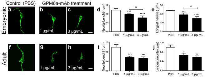
GPM6a is Involved in Neurite Outgrowth in Rat Dorsal Root Ganglion Neurons
Dorsal root ganglion (DRG) neurons regulate the expression of different molecules, such as neurotrophins and their receptors, to promote axon regeneration after injury. The membrane glycoprotein GPM6a has been described to contribute to neuronal development and structural plasticity in central-nervous-system neurons. Recent evidence indicates that GPM6a interacts with molecules from the peripheral nervous system (PNS), although its role in DRG neurons remains unknown.
To study if GPM6a contributes to neurite outgrowth in peripheral sensory neurons, embryonic and adult DRG dissociated neurons were treated with neutralizing monoclonal antibodies against GPM6a extracellular domains. Figure 1 shows representative images of control neurons treated with PBS (Figure 1a,f), or experimental neurons treated with 1 µg/mL of GPM6a-mAb (Figure 1b,g) or 3 µg/mL of GPM6a-mAb (Figure 1c, h) for 24 h. Figure 1d-e,i,j show the quantitative image analysis of the average neurite length and the longest distance reached by sensory neurites, showing a significant decrease in neurons treated with GPM6a-mAb. In agreement with what was documented for the CNS neurons, here we demonstrate that GPM6a participates in peripheral sensory neuron development and axon regeneration in vitro.
Ask a Question
Write your own review
- You May Also Need

Description: The thoracic aorta is located in the chest cavity and gives off arteries that branch to the esophagus, pericardium, lungs, and trachea. The thoracic aorta can be subdivided into the ascending aorta, ...

Description: Rat Podocytes are isolated from normal rat kidney. The cells are characterized by immunofluorescence with antibodies specific to podocin, Ang1, Nephrin, ACTN4, NPHS2. T25 flasks is required for cell ...

Description: Rat Bronchial Smooth Muscle Cells are isolated from normal rat bronchi tissue. Rat Bronchial Smooth Muscle Cells are characterized by immunofluorescence with antibodies specific to alpha-actin. T25 ...

Description: Guinea Pig Endothelial Cells from Creative Bioarray are isolated from guinea pig tissue. Prior to shipping, cells at passage 2 are detached from flasks and immediately cryopreserved in vials. Each ...

Description: Rat Lung Epithelial Cells are isolated from normal rat lung tissue. The cells are characterized by immunofluorescence with antibodies specific to CK-18, CK-19. T25 flasks is required for cell ...

Description: Rat Vein Endothelial Cells from Creative Bioarray are isolated from inferior vena cava tissue of 6-8 week old laboratory Sprague-Dawley rat. Rat Vein Endothelial Cells are grown in T75 tissue culture ...