
Human Dopaminergic Neuronal Precursor Cell
Cat.No.: CSC-C9341W
Species: Human
Source: Brain
Morphology: Multipolar
Cell Type: Neuron
- Specification
- Background
- Scientific Data
- Q & A
- Customer Review
Human Dopaminergic Neuronal Precursor Cells (hDNPs) are lineage-restricted neural progenitors that differentiate into dopamine-producing neurons of the midbrain. These cells are often derived from human pluripotent stem cells (hESCs or hiPSCs) or fetal neural tissues and represent an important intermediate stage between neural stem cells and mature dopaminergic neurons. hDNPs typically express early neuronal and dopaminergic lineage markers, like Nestin, SOX2, LMX1A, FOXA2, and NURR1, and gradually gain the ability to express tyrosine hydroxylase (TH), the rate-limiting enzyme for dopamine synthesis.
Functionally, hDNPs exhibit high proliferative capacity and can be efficiently induced to differentiate into mature, electrically active dopaminergic neurons under defined culture conditions. Upon differentiation, these neurons exhibit characteristic morphology, elaborate complex neurites, and synthesize and release dopamine. hDNPs are widely used as in vitro models to study midbrain development, neuronal differentiation and dopaminergic lineage specification because of their developmental relevance. These cells are especially valuable for studying neurodegenerative diseases, like Parkinson's, where the loss of dopaminergic neurons is a key feature. They are also used in drug screening, neurotoxicity testing and cell replacement therapy research providing a promising platform for regenerative medicine and CNS drug discovery.
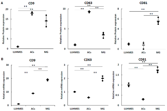
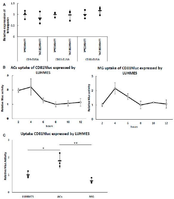
Uptake of LUHMES-Derived EVs by Astrocytes (ACs) and Microglia (MG)
Psychological stress and depression are linked to elevated interleukin-6 (IL-6), which modulates extracellular vesicle (EV) secretion. MicroRNAs (miRNAs) within EVs regulate gene expression in recipient cells upon endocytosis. Here, Nishi et al. investigated the effect of IL-6 on EVs secreted by the human dopaminergic neuronal precursor cell line (LUHMES cells).
They first characterized EV markers by measuring tetraspanin proteins in media and mRNA levels in LUHMES, astrocytes (ACs), and microglia (MG) using ELISA (Fig. 1A) and real-time PCR (Fig. 1B). While all cells expressed the three tetraspanins, no correlation was found between protein levels in the medium and cellular mRNA expression.
Next, they assessed EV uptake. Media from LUHMES cells expressing CD81-Nluc was applied to ACs and MG. Expression of CD81-Nluc did not alter endogenous tetraspanin levels in EVs (Fig. 2A), suggesting normal uptake kinetics. NanoLuc (Nluc) activity reflects the net balance of EV uptake, degradation, and recycling. Time-course analysis showed that Nluc activity in both ACs and MG peaked at 4 h and stabilized after 8 h (Fig. 2B), indicating an equilibrium state. Consequently, we compared EV uptake at the 8‑h time point. As shown in Figure 2C, ACs exhibited higher Nluc activity than MG, suggesting that astrocytes receive more EV-mediated signals from LUHMES cells than microglia.
Depending on the density of cell growth, it is routine to change the medium for 2-3 days.
Ask a Question
Average Rating: 5.0 | 1 Scientist has reviewed this product
Skilfully made
Creative Bioarray's cell products are skilfully made.
10 Sep 2023
Ease of use
After sales services
Value for money
Write your own review
Description: RFP Expressing Human Brain Microvascular Pericytes (RFP-HBMPs) provided by Creative Bioarray are zeocin-selected from Human Brain Microvascular Pericytes infected with lentivirus expressing RFP. ...
Description: Human Leptomeningeal Pericytes are isolated from normal human leptomeningeal layer which surrounds the brain in the central nervous system. They can be used in virto to study hypertension, diabetic ...

Description: GFP-Aequorin Expressing Human Brain Microvascular Endothelial Cells (GFP-AEQ-HBMVECs) provided by Creative Bioarray are puromycin-selected from Human Brain Microvascular Endothelial Cells infected ...

Description: Human Brain Vascular Adventitial Fibroblasts (HBVAF) from Creative Bioarray are isolated from human brain vascular tissue. HBVAF are cryopreserved at first passage of primary culture and delivered ...
Description: Human Primary Brain Vascular Fibroblasts are isolated from normal human brain tissue. Human Primary Brain Vascular Fibroblasts are grown in T75 tissue culture flasks pre-coated with gelatin-based ...

Description: Human dopaminergic neurons (HDNs) from Creative Bioarray are isolated from fetal brain tissue. The method we use to isolate endothelial cells was developed based on a combination of established and ...