Hamster Proximal Tubular Epithelial Cells
Cat.No.: CSC-C9207J
Species: Hamster
Source: Kidney
Cell Type: Epithelial Cell
- Specification
- Background
- Scientific Data
- Q & A
- Customer Review
Hamster proximal tubular epithelial cells (RPTECs) are primary renal epithelial cells isolated from the proximal tubules of the kidney, most often from the Syrian hamster. These cells closely resemble the physiological structure and function of the renal proximal tubule, including active reabsorption, ion transport and metabolic activity. They show characteristic epithelial morphology, form tight junctions and express major transporters, such as Na⁺/K⁺-ATPase, and markers, like megalin and aquaporins.
One of the distinctive features of hamster RPTECs is that they are derived from a hibernating species, which gives them remarkable resistance to hypoxia, oxidative stress and ischemia-reperfusion injury. Compared to rodent or human renal epithelial models, these cells exhibit greater mitochondrial stability, reduced reactive oxygen species (ROS)-induced damage, and a lessened cellular senescence response to stress. Thus, they represent a useful in vitro model to study tolerance to renal injury and adaptive metabolic regulation. Hamster RPTECs are commonly used for acute kidney injury (AKI), oxidative stress mechanisms, nephrotoxicity screening, and renal metabolism. They are also well suited to study protective signaling pathways such as the Nrf2-mediated antioxidant responses and ferroptosis resistance. In summary, these cells offer a unique and physiologically relevant platform to study kidney function and resilience to injury beyond the traditional mammalian models.
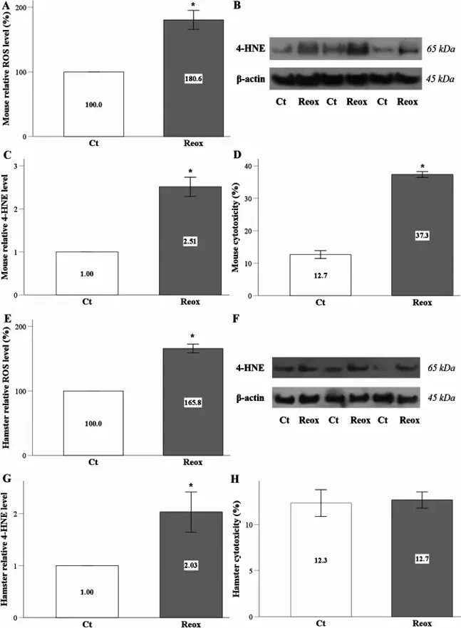
Differential Responses of RPTECs to Anoxia-Reoxygenation
Ischemia-reperfusion (I/R) injury is a leading cause of acute kidney injury (AKI), yet effective treatments are lacking. Hibernating mammals, however, show remarkable resistance to I/R-induced cell death. While cellular senescence is a key factor in AKI, its role in this context remains understudied in these species. To explore potential therapeutic insights, Pissas et al. subjected renal proximal tubular epithelial cells (RPTECs) from the hibernating Syrian hamster and the mouse to anoxia-reoxygenation.
Anoxia-reoxygenation significantly increased ROS production in mouse RPTECs (Fig. 1A) and elevated levels of 4-hydroxynonenal (4-HNE)-modified proteins, a marker of lipid peroxidation (Fig. 1B and C). Lactate dehydrogenase (LDH) release confirmed cell death in these cells (Fig. 1D). Similarly, hamster RPTECs exhibited increased ROS (Fig. 1E) and 4-HNE modification (Fig. 1F and G) following anoxia-reoxygenation. In contrast to mice, however, hamster RPTECs showed no significant cell death (Fig. 1H).
Ask a Question
Write your own review
Description: Hamster Ovarian Smooth Muscle Cells are isolated from ovarian tissue of pathogen-free laboratory mice.
Description: Hamster Esophageal Epithelial Cells from Creative Bioarray are isolated from esophageal tissue of pathogen-free laboratory mice. Hamster Esophageal Epithelial Cells are grown in a T25 tissue culture ...
Description: Hamster Spleen Epithelial Cells from Creative Bioarray are isolated from spleen tissue of pathogen-free laboratory mice. Hamster Spleen Epithelial Cells are grown in a T25 tissue culture flask ...
Description: Hamster Corneal Epithelial Cells from Creative Bioarray are isolated from corneal tissue of pathogen-free laboratory mice. Hamster Corneal Epithelial Cells are grown in a T25 tissue culture flask ...
Description: Hamster Aortic Smooth Muscle Cells are isolated from aorta of hamster.
Description: Hamster Primary Pancreatic Fibroblasts are isolated from pancreas tissue of hamster.