
Canine Uterine Epithelial Cells
Cat.No.: CSC-C9131J
Species: Dog
Source: Uterus
Cell Type: Epithelial Cell
- Specification
- Background
- Scientific Data
- Q & A
- Customer Review
Canine uterine epithelial cells are primary epithelial cells from the inner mucosal layer of the canine uterus and constitute an essential physiological and structural barrier in the female canine reproductive tract. This cell type mostly consists of endometrial luminal epithelial cells and glandular epithelial cells, which are critical for the maintenance of homeostasis of the uterine environment, embryo implantation, and gestational establishment.
They release cytokines, growth factors and uterine fluid components that affect maternal-fetal communication and immunological tolerance in pregnancy. Moreover, these cells are implicated in inflammatory responses and the pathological development of common canine reproductive illnesses, including pyometra, endometritis, and hormone-induced uterine lesions.
Canine uterine epithelial cells are widely used in in vitro experimental models to aid research in reproductive endocrinology, pathogen-host interaction, endometrial pathophysiology and drug screening for canine reproductive diseases, and also provide an important cellular tool for veterinary reproductive biology and preclinical studies.
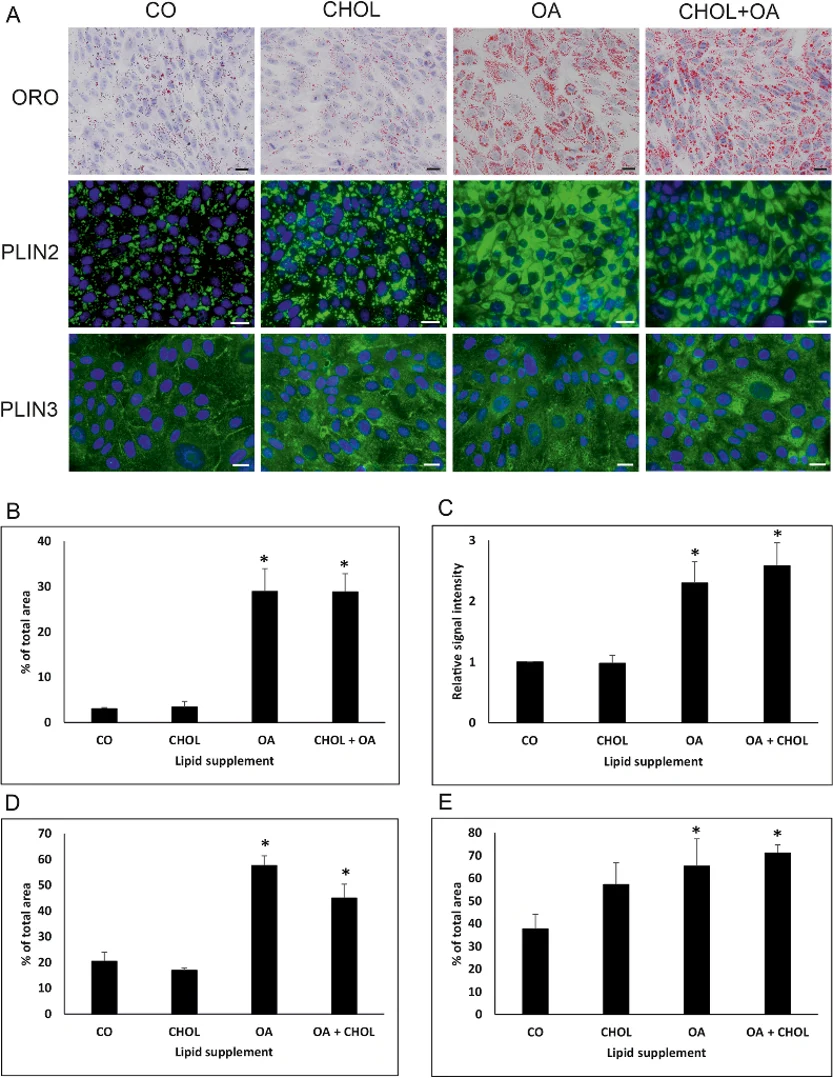
Oleic Acid Induces Lipid Droplet Accumulation in Canine Endometrial Epithelial Cells
Recent observations of lipid droplet (LD) accumulation in pyometra-affected uteri prompted a comparison of LD localization, quality, and quantity in healthy versus diseased tissues and in vitro models.
Oil Red O (ORO) staining revealed that canine endometrial epithelial cells cultured without lipid supplementation contained few, homogeneously sized LDs. However, treatment with oleic acid-alone or with cholesterol-significantly increased LD accumulation (Fig. 1A). Cholesterol alone had no effect. Quantitative image analysis showed a significant rise in ORO-positive area in oleic acid-treated groups compared to controls (Fig. 1B), which was corroborated by spectrophotometry (2-3-fold increase, Fig. 1C).
Immunostaining for perilipin 2 (PLIN2) mirrored ORO results, confirming increased LDs with oleic acid ± cholesterol treatment (Fig. 1A, D). PLIN3 staining revealed a granular cytoplasmic pattern that shifted moderately with oleic acid treatment due to enlarged LDs (Fig. 1A). Quantification showed a significant increase in PLIN3 signal in oleic acid-treated groups (Fig. 1E). These data demonstrate that canine endometrial epithelial cells actively process exogenous fatty acids, leading to LD accumulation.
Ask a Question
Write your own review
Description: Dog Liver Endothelial Cells from Creative Bioarray are isolated from tissue of dog liver. Dog Liver Endothelial Cells are grown in T25 tissue culture flasks pre-coated with gelatin-based coating ...
Description: Canine Astrocytes from Creative Bioarray are isolated from canine brain tissue. The method we use to isolate canine astrocytes were developed based on a combination of established and our proprietary ...
Description: Canine Mammary Microvascular Endothelial Cells from Creative Bioarray are isolated from breast of pathogen-free laboratory Canine. Canine Mammary Microvascular Endothelial Cells are grown in T25 ...
Description: Canine Chondrocytes (CnC) provided by Creative Bioarray are isolated from normal canine articular cartilage tissue. The cells are frozen at passage 1 and each vial contains at least 0.5*10^6 cells. ...
Description: Canine Pancreatic Microvascular Endothelial Cells from Creative Bioarray are isolated from Pancreatic Microvascular of pathogen-free laboratory Canine. Canine Pancreatic Microvascular Endothelial ...
Description: Canine Prostate Microvascular Endothelial Cells from Creative Bioarray are isolated from prostate of pathogen-free laboratory Canine. Canine Prostate Microvascular Endothelial Cells are grown in T25 ...