
Rabbit Schwann Cells
Cat.No.: CSC-C5220S
Species: Rabbit
Source: Brain
Cell Type: Schwann Cell; Glial Cell
- Specification
- Background
- Scientific Data
- Q & A
- Customer Review
Rabbit Schwann cells from Creative Bioarray are isolated from the rabbit brain tissue. The method we use to isolate rabbit Schwann cells was developed based on a combination of established and our proprietary methods. The rabbit Schwann cells are characterized by immunofluorescence with antibodies specific to glial fibrillary acidic protein(GFAP). Each vial contains 0.5x10^6 cells per ml and is delivered frozen.
Rabbit Schwann Cells are the major glial cells obtained from the peripheral nervous system of rabbits. Sciatic or spinal nerves are the most common source. They are the major myelinating cells in the PNS, wrapping axons in order to promote quick saltatory conduction and to offer critical neurotrophic support for neuronal survival and regeneration.
In vitro these cells have a bipolar or tripolar spindle-shaped morphology and develop as adherent monolayers. To retain their phenotype and prevent fibroblast expansion, they need a specialized basal media supplemented with mitogens like heregulin or forskolin. Standard culture conditions are incubation at 37°C in a humidified environment of 5% carbon dioxide.
These cells are an important model for the study of peripheral nerve biology, disorders of demyelination (such as Guillain-Barré syndrome) and repair of nerve injury. They are especially useful in the field of neuroregeneration research where they are employed for the assessment of axonal guidance, myelination processes and biocompatibility of nerve conduits or tissue-engineered scaffolds. In addition, rabbit Schwann cells are a valuable comparative model for preclinical studies spanning from rodents to human therapeutic applications.
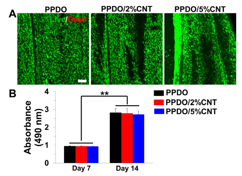
PPDO/CNT NYs Supported rSCs Growth, and Proliferation
To overcome the limitations of nerve guidance conduits (NGCs), electrically conductive PPDO/CNT composite nanofiber yarns were fabricated. Characterization confirmed successful CNT incorporation without altering the fibrous structure.
Rabbit Schwann cells (rSCs) were grown on PPDO and PPDO/CNT yarns for a period of 14 days. Live/Dead assays revealed high cell viability across all groups, with negligible cell death (Fig. 1A). Contact guidance was observed in cells orientated parallel to the nanofiber orientation (Fig. 1A). In the MTT experiment, cell counts increased significantly from day 7 to day 14, but no significant changes were observed in the proliferation rates between the PPDO and PPDO/CNT groups (Fig. 1B). These findings demonstrate that the inclusion of CNTs keeps high biocompatibility and allows cell growth and alignment, which suggests that these yarns are good scaffolds for brain tissue engineering.
Ask a Question
Write your own review
Description: Rabbit Hepatocytes are derived from the liver of New Zealand White Rabbit.
Description: The aortic arch is the top part of the main artery carrying blood away from the heart. It is the connection between the ascending and descending aorta, and its central part is formed by the left 4th ...

Description: The synovium secretes synovial fluid, which plays an important role in joint movement. The normal synovium has two layers, a thin cellular layer (luminal layer) and a vascular layer (subintima). ...
Description: The pituitary gland is an important endocrine gland in the body that secretes growth hormone and adrenocorticotropic hormone. It plays an important role in the growth and development of the body, ...
Description: The oral epthelial cells are responsible for important functions, like the primary protection of oral mucosa against external aggressions building a mechanical barrier against microorganisms, ...
Description: The carotid arteries are major blood vessels in the neck that supply blood to the brain, neck, and face. There are two carotid arteries, one on the right and one on the left. In the neck, each ...