Rabbit Peritoneal Macrophages
Cat.No.: CSC-C5273S
Species: Rabbit
Source: Peritoneal Cavity
Cell Type: Macrophage
- Specification
- Background
- Scientific Data
- Q & A
- Customer Review
Rabbit peritoneal macrophages from Creative Bioarray are isolated from the rabbit abdominal cavity. The method we use to isolate rabbit peritoneal macrophages was developed based on a combination of established and our proprietary methods. The rabbit peritoneal macrophages are characterized by immunofluorescence with antibodies specific to CD68. Each vial contains 0.5x10^6 cells per ml and is delivered frozen.
Rabbit Peritoneal Macrophages (RPMs) are primary innate immune cells extracted from the peritoneal cavity of healthy rabbits, either as resident populations or generated by inflammatory agents such as thioglycollate broth. These highly specialized phagocytic cells have characteristic macrophage morphology, with an irregular shape, strong adherence, and ample cytoplasm, while expressing classical macrophage markers such as CD68, CD11b, F4/80, iNOS, and MHC-II.
These cells preserve essential physiological capabilities, including phagocytosis, antigen presentation, and the release of inflammatory mediators such as TNF‑α, IL‑1, IL‑6, and anti‑inflammatory cytokines. RPMs serve as a dependable and extensively utilized in vitro model for immunological research, inflammatory modulation, host defense against microbial infections, and pharmacological screening. Despite constraints imposed by finite survival duration, donor-dependent variability, and the inability for prolonged passage, their vigorous immune activity and strong correlation to mammalian innate immunity render them essential for investigations into macrophage polarization, oxidative stress, autophagy, and the assessment of anti-inflammatory pharmaceuticals.
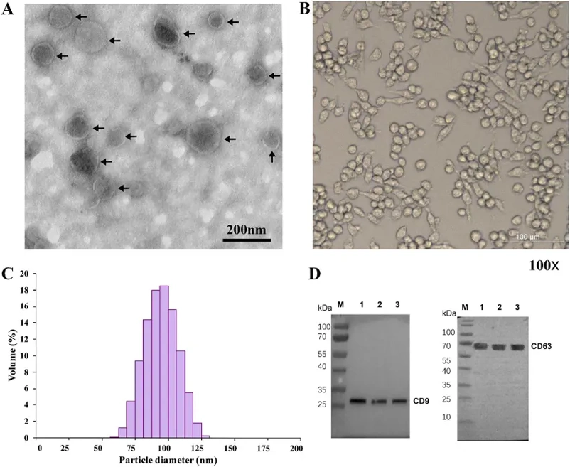
Uptake of T. pisiformis Cysticercus Exosomes by Rabbit Peritoneal Macrophages
Taenia pisiformis cysticercus infection is common in lagomorphs, causing serious economic losses in rabbit breeding. The parasite employs various immune evasion strategies, including exosome release, though the mechanism for long-term survival remains unclear. Using small RNA sequencing and TMT labeling proteomics, Wang et al. profiled miRNA and protein expression in rabbit peritoneal macrophages treated with T. pisiformis exosomes.
TEM and NTA were used to confirm the morphology and size of T. pisiformis cysticercus exosomes. The results showed typical cup-shaped vesicle structures with a diameter of 50-150 nm (Fig. 1A and Fig. 1C). Western blot confirmed the presence of exosome markers CD9 and CD63 (Fig. 1D). Additionally, rabbit peritoneal macrophages exhibited typical morphological features (round, oval, or irregular; Fig. 1B). These results confirmed successful isolation of exosomes and macrophages.
To verify whether T. pisiformis cysticercus-derived exosomes can be taken up by host cells, DiD-labeled exosomes were co-incubated with rabbit peritoneal macrophages, and fluorescence microscopy was used to track their entry. Compared with the control, red-fluorescent DiD-labeled exosomes were localized in macrophage cytoplasm (Fig. 2), indicating active uptake of exosomes by macrophages.

Ask a Question
Write your own review

Description: Rabbit Hepatocytes are derived from the liver of New Zealand White Rabbit.
Description: The aortic arch is the top part of the main artery carrying blood away from the heart. It is the connection between the ascending and descending aorta, and its central part is formed by the left 4th ...
Description: The synovium secretes synovial fluid, which plays an important role in joint movement. The normal synovium has two layers, a thin cellular layer (luminal layer) and a vascular layer (subintima). ...
Description: The pituitary gland is an important endocrine gland in the body that secretes growth hormone and adrenocorticotropic hormone. It plays an important role in the growth and development of the body, ...
Description: The oral epthelial cells are responsible for important functions, like the primary protection of oral mucosa against external aggressions building a mechanical barrier against microorganisms, ...
Description: The carotid arteries are major blood vessels in the neck that supply blood to the brain, neck, and face. There are two carotid arteries, one on the right and one on the left. In the neck, each ...