

Rabbit Corneal Endothelial Cells
Cat.No.: CSC-C5252S
Species: Rabbit
Source: Cornea; Eye
Cell Type: Endothelial Cell
- Specification
- Background
- Scientific Data
- Q & A
- Customer Review
Rabbit corneal endothelial cells from Creative Bioarray are isolated from the rabbit eye tissue. The method we use to isolate corneal endothelial cells was developed based on a combination of established and our proprietary methods. The rabbit corneal endothelial cells are characterized by immunofluorescence with antibodies specific to pan-cytokeratin (PCK). Each vial contains 0.5x10^6 cells per ml and is delivered frozen.
Rabbit corneal endothelial cells (RCECs) form the single layer of neuroectoderm-derived cells lining the posterior corneal surface. A principal strength of RCECs is their ability to establish highly reproducible, contact-inhibited monolayers exhibiting the classic polygonal, predominantly hexagonal morphology. These cells develop well-organized apicolateral tight junctions, demarcated by continuous ZO-1 localization, and uniformly express the defining endothelial markers Na⁺/K⁺-ATPase, aquaporin-1, and N-cadherin, while retaining robust transendothelial electrical resistance. Crucially, unlike primary human corneal endothelial cells, which are notoriously post-mitotic and challenging to expand, RCECs—especially those derived from young rabbits—retain a marked intrinsic proliferative capacity. This permits transient expansion without immediate loss of phenotype, enabling mechanistic studies of cell cycle re-entry and regenerative repair that are difficult in human cells.
The rabbit origin confers substantial practical and translational benefits. The large eye size simplifies endothelial isolation, yielding abundant, highly pure cells free from stromal contamination. The controlled age and genetic uniformity of laboratory rabbits minimize donor variability, ensuring experimental consistency. Furthermore, the direct alignment with established rabbit models of corneal endothelial injury, cryoinjury, and Descemet membrane transplantation enables seamless in vitro–in vivo correlation. Together, these features establish primary RCECs as a cost-effective, ethically accessible, and physiologically faithful platform for investigating endothelial pathophysiology, drug toxicity, and tissue-engineering strategies.
IGFBP4 Suppresses EndMT by Inhibiting the Wnt2/β-Catenin Signaling Pathway in Rabbit Corneal Endothelial Cells
Endothelial-mesenchymal transition (EndMT) represents a major limiting factor in the in vitro expansion of corneal endothelial cells (CECs). This study aims to identify the key genes and signaling pathways driving EndMT in rabbit CECs (RCECs) and to explore targeted interventions to preserve cellular phenotype, ultimately improving in vitro culture protocols.
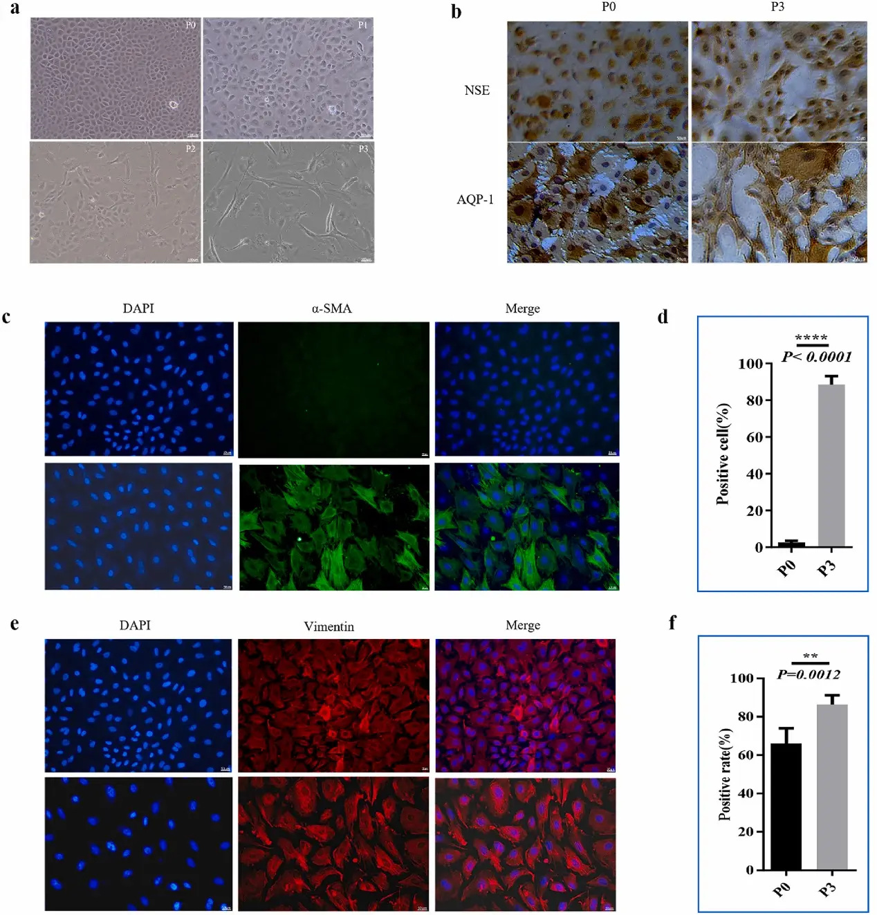
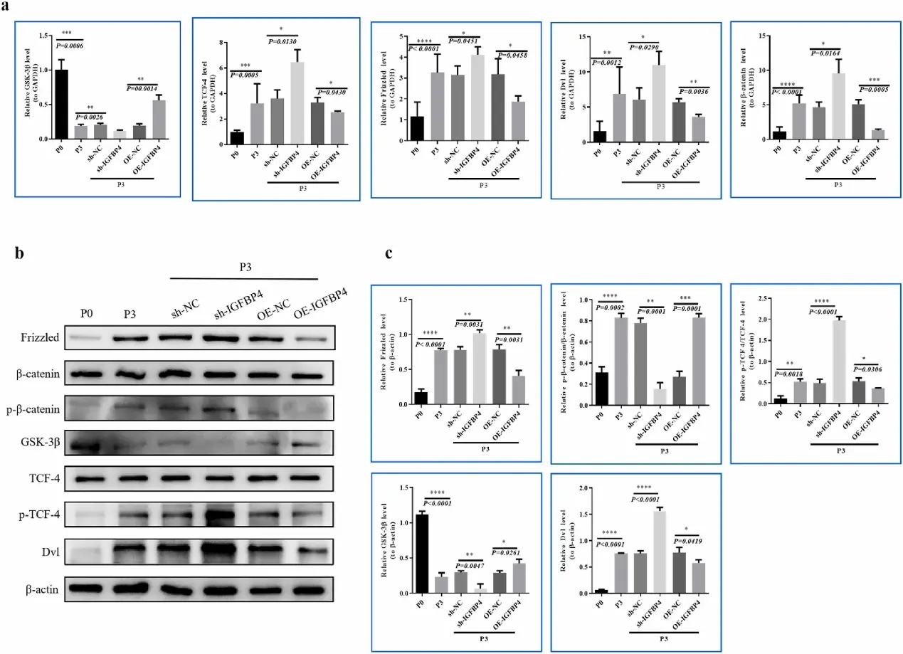
During serial passaging, primary RCECs underwent significant EndMT, characterized by progressive cellular enlargement and the acquisition of a fibroblast-like morphology. Comparative transcriptome analysis via high-throughput RNA sequencing of passage 0 (P0) and passage 3 (P3) RCECs revealed significant differential expression of EndMT-associated genes. Specifically, bioinformatic analyses (GO/KEGG) highlighted the downregulation of IGFBP4 and the upregulation of WNT components, implicating Wnt pathway activation in the EndMT process. Functionally, overexpressing IGFBP4 or knocking down WNT2 in RCECs substantially decreased the expression of EndMT markers (α-SMA and vimentin) as well as core components of the Wnt2/β-catenin pathway (Frizzled, Dvl, p-β-catenin, and p-TCF-4). Conversely, opposite genetic manipulations yielded elevated expression levels. Collectively, these findings demonstrate that IGFBP4 effectively suppresses EndMT and maintains the functional CEC phenotype by inhibiting the Wnt2/β-catenin signaling pathway.
Ask a Question
Write your own review
- You May Also Need
Description: Rabbit Hepatocytes are derived from the liver of New Zealand White Rabbit.
Description: The aortic arch is the top part of the main artery carrying blood away from the heart. It is the connection between the ascending and descending aorta, and its central part is formed by the left 4th ...
Description: The synovium secretes synovial fluid, which plays an important role in joint movement. The normal synovium has two layers, a thin cellular layer (luminal layer) and a vascular layer (subintima). ...
Description: The pituitary gland is an important endocrine gland in the body that secretes growth hormone and adrenocorticotropic hormone. It plays an important role in the growth and development of the body, ...
Description: The oral epthelial cells are responsible for important functions, like the primary protection of oral mucosa against external aggressions building a mechanical barrier against microorganisms, ...
Description: The carotid arteries are major blood vessels in the neck that supply blood to the brain, neck, and face. There are two carotid arteries, one on the right and one on the left. In the neck, each ...