

Mouse Renal Tubular Epithelial Cells
Cat.No.: CSC-C4744Z
Species: Mouse
Source: Kidney
Cell Type: Epithelial Cell
- Specification
- Background
- Scientific Data
- Q & A
- Customer Review
Never can cryopreserved cells be kept at -20 °C
Mouse renal tubular epithelial cells (mRTECs) are primary cells isolated from the mouse renal cortex. Primary mRTECs are routinely identified by positive immunofluorescence for cytokeratin 18 (CK-18), and express proximal tubule-specific markers including villin, aquaporin 1 (AQP1), and sodium-glucose cotransporter 2 (SGLT2).
The foremost advantage of mRTECs lies in their preservation of in vivo-like differentiated functions, a feature lost in immortalized cell lines. Confluent cultures form polarized monolayers with tight junctions and intact transcellular transport, as evidenced by dome formation. These cells retain proximal tubule-specific functional properties, including parathyroid hormone (PTH)-stimulated cAMP synthesis, 24,25-dihydroxyvitamin D3 production, high alkaline phosphatase activity, and Na+-dependent phosphate and α-methylglucoside transport with kinetic parameters matching brush border membrane vesicles. mRTECs can be subcultured for approximately 4-5 passages while maintaining high energetic status and mitochondrial function, making them amenable to continuous ex vivo studies.
mRTECs serve as an indispensable platform for investigating the pathophysiology of acute kidney injury, chronic kidney disease, renal fibrosis, and ischemic injury. Their compatibility with genetically modified mouse strains enables the study of cell-autonomous gene functions through transgenesis or AAV-mediated gene delivery prior to isolation. Furthermore, mRTECs are widely utilized for high-throughput drug screening, toxicological assessment, and mitochondrial bioenergetics analysis. Despite their limited proliferative capacity and propensity for phenotypic drift upon prolonged passage, optimized culture conditions-including serum-free media and matrix-independent growth-support their functional integrity, positioning mRTECs as a gold-standard ex vivo model for renal research.
Exosomes Secreted by Ferroptotic Renal Tubular Epithelial Cells Promote M1/M2 Ratio Imbalance
Severe renal ischemia and reperfusion injury (IRI) progresses to renal interstitial fibrosis (RIF) with limited therapeutic strategies. Although ferrptosis and macrophage polarization both play important roles in this model, their specific pathogenesis and interactions have not been elucidated. Therefore, we aimed to explore the mechanisms by which ferrotosis occurs in renal tubular epithelial cells (RTECs) and ferroptotic cell-derived exosomes induce macrophage polarization in IRI-related RIF model.
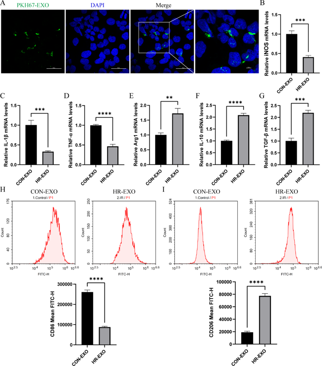
In vitro, RTECs were divided into control (CON) group, hypoxia/reoxygenation (HR) group, HR + Ferrostatin-1 (Fer-1) group, HR + siRNA-ATF3 (siATF3) group. Subsequently, we co-cultured exosomes derived from CON or HR groups with RAW264.7 cells (CON-EXO or HR-EXO). Intracellular transport of the exosomes into RAW264.7 cells was shown in Figure 1A, as PKH67-labeled exosomes (green fluorescence) were taken up by RAW264.7 cells and colocalized with DAPI-labeled nuclei (blue fluorescence). We detected the concentration of M1 markers (iNOS, IL-1β, and TNF-α) and M2 markers (Arg1, IL-10, and TGF-β) in the culture supernatant, and found that exosomes inhibited the production of iNOS, IL-1β, and TNF-α (Figures 1B-D), and enhanced Arg1, IL-10, and TGF-β (Figures 1E-G). Flow cytometry results also support that exosomes cause an increased proportion of CD206+ M2 phenotype macrophages (Figures 1H and I). In summary, these data indicated that exosomes secreted by ferroptotic RTECs promoted the polarization of macrophages towards M2.
Ask a Question
Write your own review
Description: C57BL/6-GFP Mouse Skeletal Muscle Microvascular Endothelial Cells from Creative Bioarray are isolated from C57BL/6-Tg (CAG-EGFP) 1Osb/J mouse skeletal muscle tissue of pathogen-free laboratory mice. ...
Description: eNOS KO Mouse Stomach Epithelial Cells from Creative Bioarray are isolated from stomach tissue of pathogen-free laboratory mice. eNOS KO Mouse Stomach Epithelial Cells are grown in a T25 tissue ...
Description: eNOS KO Mouse Liver Fibroblasts from Creative Bioarray are isolated from liver tissue of pathogen-free laboratory mice. eNOS KO Mouse Liver Fibroblasts are grown in T75 tissue culture flasks ...

Description: C57BL/6-GFP Mouse Corneal Epithelial Cells from Creative Bioarray are isolated from C57BL/6-GFP-Tg(CAG-EGFP)1Osb/J mouse corneal tissue of pathogen-free laboratory mice. C57BL/6-GFP Mouse Corneal ...
Description: BALB/c Mouse Retinal Microvascular Endothelial Cells from Creative Bioarray are isolated from retinal tissue of pathogen-free laboratory mice. BALB/c Mouse Retinal Microvascular Endothelial Cells are ...