
Mouse Renal Artery Endothelial Cells
Cat.No.: CSC-C5303S
Species: Mouse
Source: Kidney; Artery
Cell Type: Endothelial Cell
- Specification
- Background
- Scientific Data
- Q & A
- Customer Review
Mouse renal artery endothelial cells from Creative Bioarray are isolated from the mouse renal artery tissue. The method we use to isolate mouse renal artery endothelial cells was developed based on a combination of established and our proprietary methods. The mouse renal artery endothelial cells are characterized by immunofluorescence with antibodies specific to von Willebrand factor (vWF). Each vial contains 0.5x10^6 cells per ml and is delivered frozen.
Mouse Renal Artery Endothelial Cells (MRAECs) line the luminal surface of the renal artery. This endothelium is made up of a specialized layer of squamous epithelial cells that line the vascular cavity. They serve many more purposes than just lining blood vessels, however. They can actively regulate vascular tone, permeability, and renal blood flow. Endothelial cells are involved in sensing blood flow and vessel stretching and they help to regulate systemic blood pressure and pressures within the kidney.
Endothelial cells release vasodilators and vasoconstrictors to regulate smooth muscle tone. Nitric Oxide (NO) produced by endothelial Nitric Oxide Synthase (eNOS) is an important endothelial cell produced vasodilator. Mouse renal artery endothelial cells also produce vasoconstrictors such as endothelin-1 and prostaglandins. The release of these molecules maintains homeostasis by preventing excess vasoconstriction of the renal blood vessels. Endothelial cells maintain homeostasis of other mechanisms as well. Mouse renal artery endothelial cells can control coagulation within the vessel lumen. In addition, endothelial cells help regulate leukocyte binding to the endothelial layer by expressing molecules such as ICAM-1 and VCAM-1 during inflammation. An example of when things go wrong in the endothelium is endothelial dysfunction. Endothelial dysfunction can lead to many severe diseases.
Mouse renal artery endothelial cells are often used in cardiovascular and urological research models. These models include Renovascular Hypertension, Atherosclerosis, and Acute Kidney Injury (AKI). Since the renal artery supplies oxygenated blood to the nephrons, anything that damages the endothelial cells can cause ischemia and kidney fibrosis downstream.
Cytotoxicity of Hydrophobic Chitosan/Salicylic Acid Blends Film
Degradable plastic materials made from biopolymer chitosan (CS) are potential alternatives to plastic bags for food packaging, but their stability and durability are limited. Liu's team demonstrated a novel method to produce hydrophobic CS/salicylic acid (SA) films in an organic phase, aiming to improve their properties for food packaging applications.
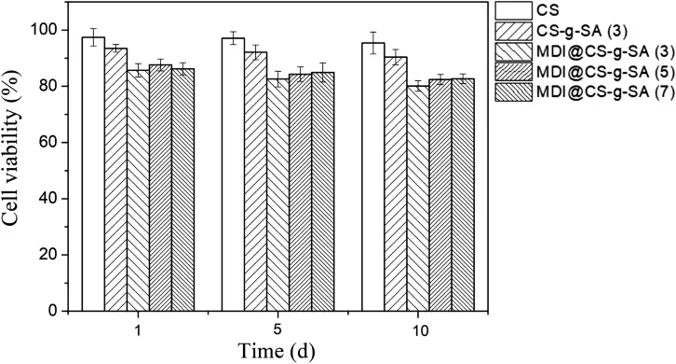
The cytotoxicity of MDI crosslinked films was assessed using an MTT assay with mouse renal artery endothelial cells. The cytotoxicity of MDI crosslinked film is an important index of food packaging materials. In this study, they used the MTT test to prove the cytotoxicity of the MDI crosslinked CS films. As shown in Figure 1, the MTT assay shows that MDI crosslinked films still exhibited low inhibitory effect on the cell viability within 10 days. The value of all the cell viability exceeds 80%, indicating MDI crosslinked films is safe to use in food packaging.
Ask a Question
Write your own review
- You May Also Need

Description: C57BL/6-GFP Mouse Skeletal Muscle Microvascular Endothelial Cells from Creative Bioarray are isolated from C57BL/6-Tg (CAG-EGFP) 1Osb/J mouse skeletal muscle tissue of pathogen-free laboratory mice. ...

Description: eNOS KO Mouse Stomach Epithelial Cells from Creative Bioarray are isolated from stomach tissue of pathogen-free laboratory mice. eNOS KO Mouse Stomach Epithelial Cells are grown in a T25 tissue ...

Description: eNOS KO Mouse Liver Fibroblasts from Creative Bioarray are isolated from liver tissue of pathogen-free laboratory mice. eNOS KO Mouse Liver Fibroblasts are grown in T75 tissue culture flasks ...

Description: C57BL/6-GFP Mouse Corneal Epithelial Cells from Creative Bioarray are isolated from C57BL/6-GFP-Tg(CAG-EGFP)1Osb/J mouse corneal tissue of pathogen-free laboratory mice. C57BL/6-GFP Mouse Corneal ...

Description: BALB/c Mouse Retinal Microvascular Endothelial Cells from Creative Bioarray are isolated from retinal tissue of pathogen-free laboratory mice. BALB/c Mouse Retinal Microvascular Endothelial Cells are ...