
Mouse Middle Ear Epithelial Cells
Cat.No.: CSC-C5402S
Species: Mouse
Source: Ear
Cell Type: Epithelial Cell
- Specification
- Background
- Scientific Data
- Q & A
- Customer Review
Mouse middle ear epithelial cells from Creative Bioarray are isolated from the mouse middle ear tissue. The method we use to isolate mouse middle ear epithelial cells was developed based on a combination of established and our proprietary methods. The mouse middle ear epithelial cells are characterized by immunofluorescence with antibodies specific to pan-cytokeratin (PCK). Each vial contains 0.5x10^6 cells per ml and is delivered frozen.
Mouse Middle Ear Epithelial Cells Mouse middle ear epithelial cells are primary cells isolated from the mucosal lining of the mouse middle ear cavity. This tissue is similar to respiratory epithelium and contains ciliated, secretory and basal cell populations important for middle ear homeostasis, sterility and mucociliary clearance. In vitro, these cells show a typical epithelial morphology, forming adherent monolayers of polygonal or cobblestone-like cells. They are usually maintained in a basal medium supplemented with nutrients under standard conditions (37 °C, 5% CO2). They have a short proliferative lifespan which makes them delicate to handle and are often cultured with specialized supplements to maintain their differentiated phenotype.
These cells constitute a physiologically relevant model and are thus indispensable in otological research, particularly for the study of the pathogenesis of otitis media (middle ear inflammation) and cholesteatoma. They permit the study of epithelial remodeling, mucus hypersecretion and innate immune responses to otopathogens such as Haemophilus influenzae. They also provide an important platform for evaluating drug efficacy, assessing middle ear barrier function and studying host-pathogen interactions in the auditory system.
Transcriptomic Profiling of Mouse Middle Ear Epithelial Cells During Mucociliary Differentiation
Otitis media (OM) is a prevalent pediatric disease driven by epithelial dysfunction, yet the mechanisms of middle ear remodeling are poorly understood due to limited model systems. Mulay et al. utilized a novel in vitro model of mouse middle ear epithelial cells (mMEECs) capable of mucociliary differentiation to characterize genome-wide transcriptional changes during maturation.
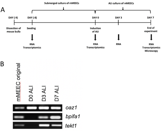
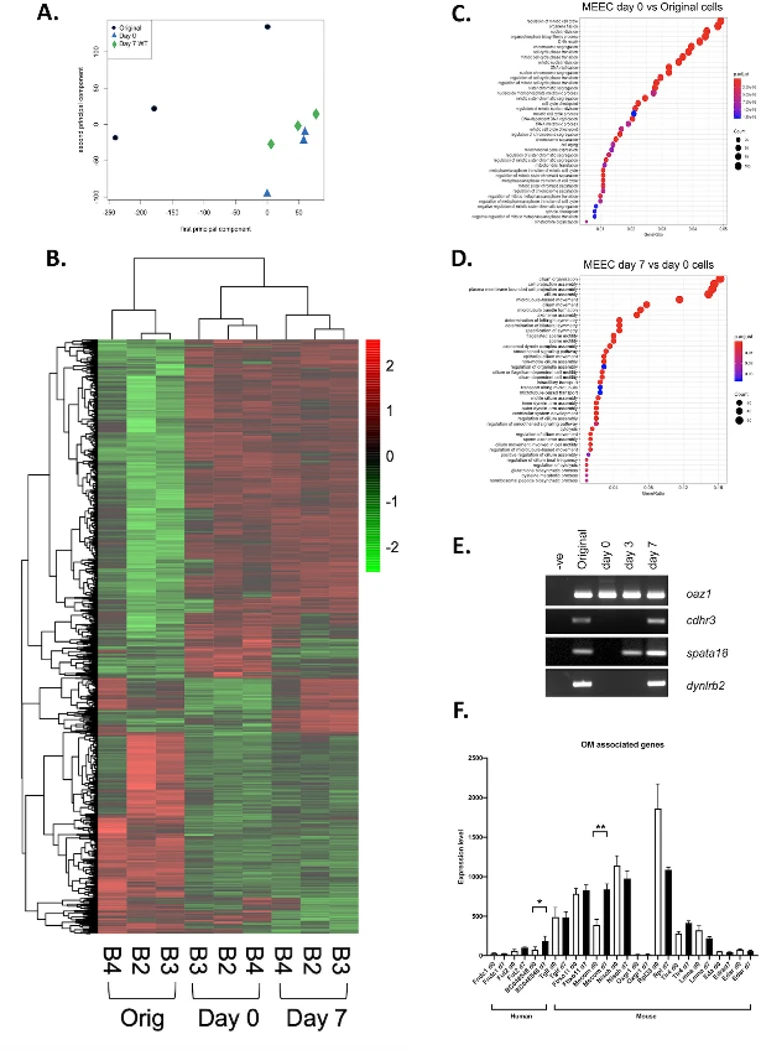
mMEECs were cultured at an air-liquid interface (ALI) for 7 days (Fig. 1A). End-point PCR confirmed the upregulation of differentiation markers for secretory (Bpifa1) and ciliated (Tekt1) cells within 3 days (Fig. 1B). Gene expression arrays compared original (uncultured) cells, undifferentiated confluent cells (Day 0 ALI), and differentiated ALI cells (Day 7). Original cells exhibited a mixed population with high expression of hemoglobin and secretory proteins (Bpifa1, Lyz2, Bpifb1). By Day 0, expression shifted to structural genes (e.g., Lcn2). By Day 7 ALI, the profile was dominated by secretory proteins (Lcn2, Reg3g, Bpifa1, Cp, Ltf, Tf), aligning with prior proteomic data. Principal Component Analysis confirmed distinct clustering of the three groups, with original cells showing the greatest variability (Fig. 2A).
Ask a Question
Write your own review
- You May Also Need


Description: C57BL/6-GFP Mouse Skeletal Muscle Microvascular Endothelial Cells from Creative Bioarray are isolated from C57BL/6-Tg (CAG-EGFP) 1Osb/J mouse skeletal muscle tissue of pathogen-free laboratory mice. ...

Description: eNOS KO Mouse Stomach Epithelial Cells from Creative Bioarray are isolated from stomach tissue of pathogen-free laboratory mice. eNOS KO Mouse Stomach Epithelial Cells are grown in a T25 tissue ...

Description: eNOS KO Mouse Liver Fibroblasts from Creative Bioarray are isolated from liver tissue of pathogen-free laboratory mice. eNOS KO Mouse Liver Fibroblasts are grown in T75 tissue culture flasks ...

Description: C57BL/6-GFP Mouse Corneal Epithelial Cells from Creative Bioarray are isolated from C57BL/6-GFP-Tg(CAG-EGFP)1Osb/J mouse corneal tissue of pathogen-free laboratory mice. C57BL/6-GFP Mouse Corneal ...

Description: BALB/c Mouse Retinal Microvascular Endothelial Cells from Creative Bioarray are isolated from retinal tissue of pathogen-free laboratory mice. BALB/c Mouse Retinal Microvascular Endothelial Cells are ...