Mouse Gallbladder Epithelial Cells
Cat.No.: CSC-C5307S
Species: Mouse
Source: Gallbladder
Cell Type: Epithelial Cell
- Specification
- Background
- Scientific Data
- Q & A
- Customer Review
Mouse gallbladder epithelial cells from Creative Bioarray are isolated from the mouse gallbladder tissue. The method we use to isolate mouse gallbladder epithelial cells was developed based on a combination of established and our proprietary methods. The mouse gallbladder epithelial cells are characterized by immunofluorescence with antibodies specific to cytokeratin-19 (CK-19). Each vial contains 0.5x10^6 cells per ml and is delivered frozen.
Mouse gallbladder epithelial cells (mGBECs) are primary epithelial cells isolated from the mucosal surface of the mouse gallbladder, which is a small bile duct-like organ that concentrates, stores and secretes bile. Mouse GBECs are typically derived from C57BL/6 mice and display cuboidal to columnar morphology, have tight junctions and express epithelial markers such as CK-19, EpCAM and CD49f, while lacking expression of endothelial (CD31) and immune cell markers (CD45). These cells function to absorb water/electrolytes from bile, secrete mucus and regulate homeostasis of the biliary barrier. mGBECs retain high proliferative potential in cell culture and differentiate into hepatocyte-like cells when induced. mGBECs are one of the most commonly utilized cell models for studying biliary biology and diseases. Current models include gallstone disease, cholecystitis, biliary injury repair, and liver regeneration to study EMT and stem cell mediated repair.
Insulin Expression in Developing Mouse Gallbladder Cells
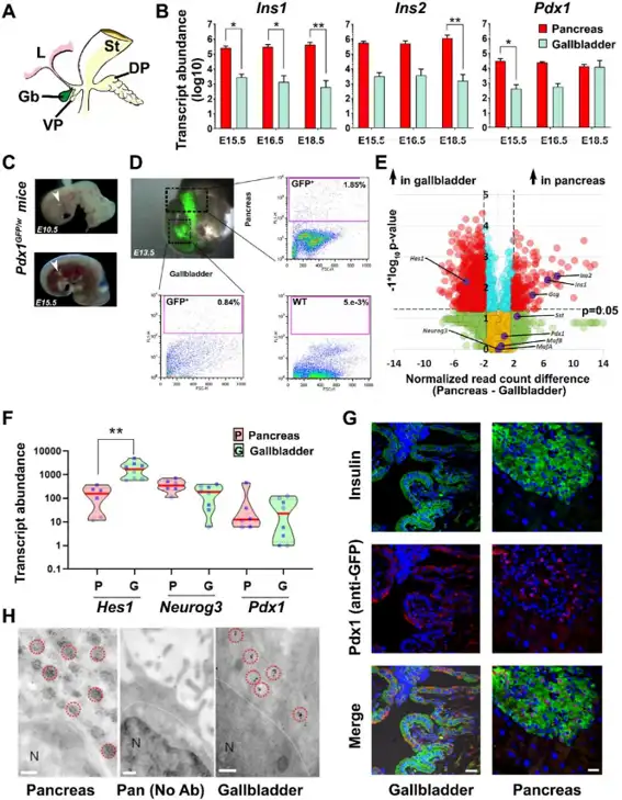
Pancreatic islet β-cells are the primary insulin source, though ectopic expression is recognized. Given the gallbladder's developmental proximity to the pancreas, Joglekar et al. investigated whether gallbladders contain functional insulin-producing cells using immunohistochemistry, flow cytometry, ELISAs, RNA-seq, qPCR, ChIP, and functional assays in mouse and human tissues.
The gallbladder originates from the pancreatic bud (Fig. 1A). During development (E15.5-E18.5), pancreas contained higher Ins1/Ins2 transcripts, but Pdx1 was expressed in gallbladder at comparable levels near birth (Fig. 1B); other transcripts showed no difference. Gallbladders contained immunoreactive insulin at lower levels. Pdx1GFP/w mice confirmed Pdx1 promoter activity in gallbladder buds (Fig. 1C, D), with ChIP verifying active promoter sites.
Adult RNA-seq showed higher pancreatic hormones in pancreas, but elevated Hes1 (Neurog3 repressor) in gallbladder (Fig. 1E). Pdx1 expression remained similar (Fig. 1E, F), with lower Neurog3 and higher Hes1 in gallbladder. Pdx1GFP/w and MIP-GFP mice confirmed insulin-Pdx1 coexpression and Ins1 promoter activity in adult gallbladder (Fig. 1G). Immune-electron microscopy identified insulin secretory vesicles in mouse gallbladder epithelial cells (Fig. 1H), demonstrating pancreatic endocrine gene expression and insulin packaging capacity in mouse gallbladder.
Ask a Question
Write your own review
- You May Also Need


Description: C57BL/6-GFP Mouse Skeletal Muscle Microvascular Endothelial Cells from Creative Bioarray are isolated from C57BL/6-Tg (CAG-EGFP) 1Osb/J mouse skeletal muscle tissue of pathogen-free laboratory mice. ...

Description: eNOS KO Mouse Stomach Epithelial Cells from Creative Bioarray are isolated from stomach tissue of pathogen-free laboratory mice. eNOS KO Mouse Stomach Epithelial Cells are grown in a T25 tissue ...
Description: eNOS KO Mouse Liver Fibroblasts from Creative Bioarray are isolated from liver tissue of pathogen-free laboratory mice. eNOS KO Mouse Liver Fibroblasts are grown in T75 tissue culture flasks ...
Description: C57BL/6-GFP Mouse Corneal Epithelial Cells from Creative Bioarray are isolated from C57BL/6-GFP-Tg(CAG-EGFP)1Osb/J mouse corneal tissue of pathogen-free laboratory mice. C57BL/6-GFP Mouse Corneal ...

Description: BALB/c Mouse Retinal Microvascular Endothelial Cells from Creative Bioarray are isolated from retinal tissue of pathogen-free laboratory mice. BALB/c Mouse Retinal Microvascular Endothelial Cells are ...