
Mouse Abdominal Aortic Adventitial Fibroblasts
Cat.No.: CSC-C5359S
Species: Mouse
Source: Aorta
Cell Type: Fibroblast
- Specification
- Background
- Scientific Data
- Q & A
- Customer Review
Mouse abdominal aortic adventitial fibroblasts from Creative Bioarray are isolated from the mouset abdominal aorta tissue. The method we use to isolate mouse abdominal aortic adventitial fibroblasts was developed based on a combination of established and our proprietary methods. The mouse abdominal aortic adventitial fibroblasts are characterized by immunofluorescence with antibodies specific to vimentin. Each vial contains 0.5x10^6 cells per ml and is delivered frozen.
Mouse abdominal aortic adventitial fibroblasts (mAAAFs) are primary vascular adventitial stromal cells originally isolated from the outermost layer (tunica adventitia) of the abdominal aorta, the largest artery located in the abdominal cavity. mAAAFs account for the majority of cells in vascular adventitia and are characterized by their spindle‑shaped morphology, their ability to adhere to plastic, and their uniform expression of fibroblast marker vimentin with concomitant lack of endothelial (vWF) and smooth muscle (desmin) markers allowing for straightforward and pure preparation of cultured cells.
Functionally, mAAAFs play an important role in vascular homeostasis, extracellular matrix remodeling and inflammation. After activation by pathological stimuli like angiotensin II or elastase injury, mAAAFs proliferate, differentiate into myofibroblasts and produce pro‑inflammatory cytokines (e.g., IL‑6, MCP‑1) and ECM proteins that promote vascular fibrosis, atherosclerosis and abdominal aortic aneurysm (AAA) development.
mAAAFs are broadly used to study adventitia‑mediated vascular remodeling and inflammation in vitro and represent an appropriate disease model to study vascular remodeling, inflammation, AAA pathogenesis and identify therapeutic targets for vascular disease.
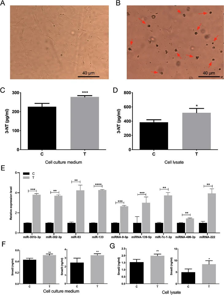
Alteration of Nitration Level and TGFβ/Smads Signaling Pathway-Related miRNA/Protein in MAFs After Acute Exposure to UFPS
The previous study showed that acute exposure to ultrafine particles (UFP, <100 nm) caused biological dysregulation in atherosclerosis. In this study, mice were exposed to UFP for 6 consecutive days followed by euthanasia at 3, 5, 7 and 10 days after exposure for collection of aorta and serum. Mouse aortic adventitial fibroblasts (MAFs) were isolated from the aorta and were utilized to study UFP uptake, oxidative stress induction, and changes in TGFβ/Smad signaling pathway.
Visualization of black dots inside the cells showed uptake of UFPs. Levels of oxidative stress marker 3-nitrotyrosine (3-NT) were significantly higher after 3 days of exposure to UFPs (Fig. 1a, b). Expression levels of several miRNAs related to TGFβ/Smads pathway were significantly upregulated by UFP exposure (miR-301b, miR-382-3p, miR-83, miR-133, miR-9-5p, miR-139-5p, let-7c-1-3p, miR-499-3p, miR-222) along with extracellular/intracellular Smad2 protein (Fig. 1e-g) and intracellular Smad3 protein in MAFs. These data indicate that UFPs accelerate progression of atherosclerosis by activating TGFβ/Smad signaling pathway.
Ask a Question
Write your own review
- You May Also Need

Description: C57BL/6-GFP Mouse Skeletal Muscle Microvascular Endothelial Cells from Creative Bioarray are isolated from C57BL/6-Tg (CAG-EGFP) 1Osb/J mouse skeletal muscle tissue of pathogen-free laboratory mice. ...

Description: eNOS KO Mouse Stomach Epithelial Cells from Creative Bioarray are isolated from stomach tissue of pathogen-free laboratory mice. eNOS KO Mouse Stomach Epithelial Cells are grown in a T25 tissue ...

Description: eNOS KO Mouse Liver Fibroblasts from Creative Bioarray are isolated from liver tissue of pathogen-free laboratory mice. eNOS KO Mouse Liver Fibroblasts are grown in T75 tissue culture flasks ...

Description: C57BL/6-GFP Mouse Corneal Epithelial Cells from Creative Bioarray are isolated from C57BL/6-GFP-Tg(CAG-EGFP)1Osb/J mouse corneal tissue of pathogen-free laboratory mice. C57BL/6-GFP Mouse Corneal ...

Description: BALB/c Mouse Retinal Microvascular Endothelial Cells from Creative Bioarray are isolated from retinal tissue of pathogen-free laboratory mice. BALB/c Mouse Retinal Microvascular Endothelial Cells are ...