

Mouse Splenocytes CD4+ T Cells
Cat.No.: CSC-C4460X
Species: Mouse
Source: Spleen
Cell Type: T Cell; Lymphocyte
- Specification
- Background
- Scientific Data
- Q & A
- Customer Review
Mouse Splenocytes CD4+ T Cells are primary CD4 positive T cells that are purified from mouse spleens. CD4+ T cells are an important aspect of the adaptive immune system and are involved in immunological control through cytokine production and interactions with other immune cell types. Once activated, these cells are capable of differentiating into various functional subsets, such as T helper 1 (Th1), T helper 2 (Th2), T helper 17 (Th17), follicular helper T (Tfh), and regulatory T (Treg) cells, which is determined by the cytokine milieu and signaling cues in the local environment.
Mouse splenic CD4+ T cells are widely employed in studies of T-cell activation, differentiation, proliferation, and cytokine release. They can be used to study antigen-specific immune responses, immunological signaling pathways, and processes of inflammation and immune control. These cells are also often used in research linked to autoimmune illnesses, infectious diseases, cancer immunology and vaccination responses.
In vitro, Mouse Splenocytes CD4+ T Cells can be activated with antibodies, antigens or cytokines to assess functional responses under defined experimental settings. They are ideal model to investigate CD4+ T-cell biology and cellular interactions in the immune system, as they are primary immune cells that originate from native lymphoid tissue. Their applications include studies of cytokine signaling, immunological regulation, differentiation of T-cell subsets, and assessment of immunotherapeutic techniques in preclinical trials.
Genetic Dissection of Eomes Function in CD4⁺ T Cells Using a Novel Tbx21ᴱ Allele
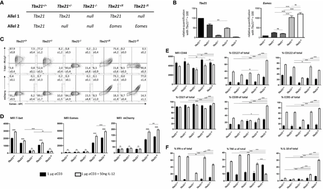
To delineate whether Eomesodermin (Eomes) mirrors or diverges from T-bet in CD4⁺ T cells, Thelen et al. employed a novel Tbx21ᴱ knock-in allele that replaces T-bet with Eomes and an mCherry reporter. Combining this with wild-type (Tbx21⁺/⁺) and knockout (Tbx21⁻/⁻) alleles generated five mouse lines with titrated T-box transcription factor expression (Fig. 1A). This design uncouples Eomes induction from chronic antigen exposure, allowing direct assessment of its lineage-specific effects under Th1-polarizing conditions.
Transcriptional profiling confirmed allele fidelity: Tbx21⁻/ᴱcells lacked Tbx21mRNA, while Tbx21⁺/ᴱand Tbx21⁻/ᴱcells exhibited robust Eomes expression (Fig. 1B). Loss of Tbx21alone caused only marginal increases in endogenous Eomes, indicating that Eomes is not a default backup for T-bet. Flow cytometry validated these findings, showing reduced T-bet protein in haploin sufficient cells and complete absence in Tbx21⁻/⁻and Tbx21⁻/ᴱ CD4⁺ T cells (Fig. 1C, 1D). Strikingly, the Tbx21ᴱ allele drove uniform, high-level Eomes expression paralleled by mCherry fluorescence, confirming that Eomes is transcribed from the engineered locus rather than the endogenous Eomes promoter. IL-12 further amplified Eomes and mCherry expression, demonstrating full transcriptional control by the Tbx21 regulatory elements. These results establish that Eomes expression from the Tbx21ᴱ allele faithfully recapitulates the dynamics of wild-type T-bet, providing a powerful genetic system to dissect Eomes-specific transcriptional programs in CD4⁺ T cells independent of environmental cues.
Ask a Question
Write your own review
Description: C57BL/6-GFP Mouse Skeletal Muscle Microvascular Endothelial Cells from Creative Bioarray are isolated from C57BL/6-Tg (CAG-EGFP) 1Osb/J mouse skeletal muscle tissue of pathogen-free laboratory mice. ...

Description: eNOS KO Mouse Stomach Epithelial Cells from Creative Bioarray are isolated from stomach tissue of pathogen-free laboratory mice. eNOS KO Mouse Stomach Epithelial Cells are grown in a T25 tissue ...
Description: eNOS KO Mouse Liver Fibroblasts from Creative Bioarray are isolated from liver tissue of pathogen-free laboratory mice. eNOS KO Mouse Liver Fibroblasts are grown in T75 tissue culture flasks ...
Description: C57BL/6-GFP Mouse Corneal Epithelial Cells from Creative Bioarray are isolated from C57BL/6-GFP-Tg(CAG-EGFP)1Osb/J mouse corneal tissue of pathogen-free laboratory mice. C57BL/6-GFP Mouse Corneal ...

Description: BALB/c Mouse Retinal Microvascular Endothelial Cells from Creative Bioarray are isolated from retinal tissue of pathogen-free laboratory mice. BALB/c Mouse Retinal Microvascular Endothelial Cells are ...