
Mouse Nephron Progenitor Cells
Cat.No.: CSC-C9367W
Species: Mouse
Source: Kidney
Morphology: Cobblestone
Cell Type: Progenitor Cell
- Specification
- Background
- Scientific Data
- Q & A
- Customer Review
Mouse Nephron Progenitor Cells (NPCs) are renal progenitor cells obtained from developing mouse kidney. During kidney development, nephron progenitor cells in the nephrogenic niche differentiate into several epithelial components of the nephron, such as podocytes, proximal tubules, distal tubules, and portions of the loop of Henle. These cells are utilized extensively to study the molecular and cellular events underlying nephrogenesis due to their developmental capacity.
These cells can be used in vitro to investigate progenitor cell maintenance, differentiation, lineage specification, and signaling pathways involved in kidney development. They are often used in studies of Wnt, FGF, BMP and Notch signaling that are critical for nephron development and progenitor cell regulation. These cells have also been used in kidney organoid systems and tissue engineering approaches to study mechanisms of renal development and morphogenesis. Mouse NPCs can be employed for research on congenital kidney diseases, developmental biology, regenerative medicine and modelling of kidney diseases. Moreover, they provide a suitable experimental system to investigate factors influencing nephron differentiation and maturation.
Long-Term Clonal Expansion of Nephron Progenitor Cells via p38 and YAP Modulation
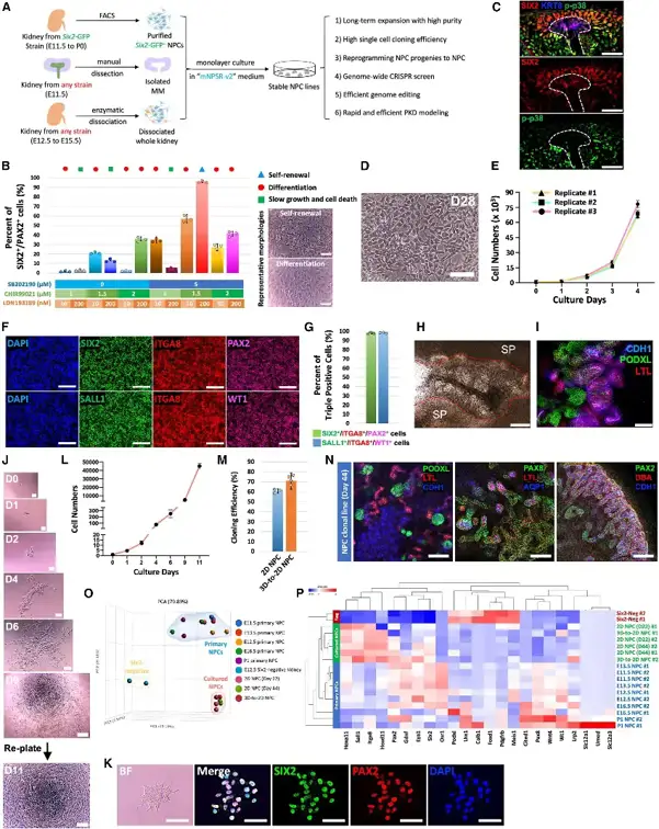
The limited expansion capacity of nephron progenitor cells (NPCs) restricts kidney research and disease modeling. To address this, Huang et al. developed mNPSR-v2, a chemically defined medium optimized for long-term clonal expansion of mouse NPCs (mNPCs) in 2D culture. Compared to the original mNPSR, mNPSR-v2 incorporates four key inhibitors: p38 MAPK (SB202190), Notch (DAPT), TGF-β (A83-01), and BMP (LDN193189), alongside adjusted CHIR99021 concentration. Among these, SB202190 was critical for maintaining the SIX2⁺/PAX2⁺ NPC pool (Fig. 1B, 1C), while DAPT prevented spontaneous differentiation.
mNPCs cultured in mNPSR-v2 proliferated stably with homogeneous morphology (Fig. 1D, 1E) and uniform expression of NPC markers (Fig. 1F, 1G). Withdrawal of any single component compromised self-renewal. Functionally, cultured mNPCs retained robust nephrogenic potential. In the spinal cord induction assay, they formed tubule-like structures containing PODXL⁺ glomeruli, LTL⁺ proximal tubules, and CDH1⁺ distal tubules (Fig. 1H, 1I). They also generated nephron organoids in defined media. drove ureteric bud branching morphogenesis, and differentiated into nephrons when transplanted in vivo.
Clonal expansion from single cells achieved 60%-70% efficiency (Fig. 1J-1N). Bulk RNA sequencing confirmed that cultured mNPCs transcriptionally resembled primary E11.5-E13.5 NPCs (Fig. 1O, 1P). This platform enabled robust derivation of mNPC lines from isolated metanephric mesenchyme or whole embryonic kidneys across multiple mouse strains, providing a scalable resource for kidney development and disease studies.
Ask a Question
Write your own review


Description: C57BL/6-GFP Mouse Skeletal Muscle Microvascular Endothelial Cells from Creative Bioarray are isolated from C57BL/6-Tg (CAG-EGFP) 1Osb/J mouse skeletal muscle tissue of pathogen-free laboratory mice. ...
Description: eNOS KO Mouse Stomach Epithelial Cells from Creative Bioarray are isolated from stomach tissue of pathogen-free laboratory mice. eNOS KO Mouse Stomach Epithelial Cells are grown in a T25 tissue ...
Description: eNOS KO Mouse Liver Fibroblasts from Creative Bioarray are isolated from liver tissue of pathogen-free laboratory mice. eNOS KO Mouse Liver Fibroblasts are grown in T75 tissue culture flasks ...

Description: C57BL/6-GFP Mouse Corneal Epithelial Cells from Creative Bioarray are isolated from C57BL/6-GFP-Tg(CAG-EGFP)1Osb/J mouse corneal tissue of pathogen-free laboratory mice. C57BL/6-GFP Mouse Corneal ...

Description: BALB/c Mouse Retinal Microvascular Endothelial Cells from Creative Bioarray are isolated from retinal tissue of pathogen-free laboratory mice. BALB/c Mouse Retinal Microvascular Endothelial Cells are ...