
Mouse Lewis Lung Tumor Endothelial Cells
Cat.No.: CSC-C8623W
Species: Mouse
Source: Lung
Cell Type: Endothelial Cell
- Specification
- Background
- Scientific Data
- Q & A
- Customer Review
Mouse Lewis Lung Tumour Endothelial Cells (LLTECs) are primary tumor-associated endothelial cells isolated from the vasculature of Lewis Lung Carcinoma (LLC) in C57BL/6 mice. Unlike normal vascular endothelial cells, LLTECs have a unique activated phenotype, which reflects the specific microenvironment of solid tumors, and are therefore an effective in vitro model to study tumor angiogenesis, vascular remodeling and tumor-stroma interactions. These cells have been shown to express a high level of endothelial markers like CD31 (PECAM-1), ICAM-2, VE-cadherin, VEGFR2 (Flk-1) and Sca-1, and to express constitutively activation-associated molecules like E-selectin, ICAM-1 and VCAM-1. These features differentiate them from normal endothelial cells, and are similar to the activated vasculature of growing tumors.
LLTECs are extensively used in cancer research to study the mechanisms of tumor vascularization, endothelial activation, leukocyte recruitment, and remodeling of the tumor microenvironment. In particular, this model is useful for the evaluation of anti-angiogenic therapies, vascular targeting agents and new drug delivery strategies, as tumor endothelial cells are critically important in the delivery of nutrients and oxygen to malignant tissues. Additionally, LLTECs provide a physiologically relevant platform to study interactions between endothelial cells, cancer cells, immune cells and extracellular matrix components.
Due to their tumor-specific phenotype and reproducible experimental performance, Mouse Lewis Lung Tumor Endothelial Cells have become an important research tool for oncology, tumor microenvironment studies, metastasis research, and the preclinical development of anti-cancer therapeutics targeting the tumor vasculature.
Whole-Exome Sequencing Reveals Distinct Mutational Landscape of Lewis Lung Carcinoma
Lewis lung carcinoma (LLC), as a widely used preclinical cancer model, has still not been genetically and genomically characterized. Here, He et al. performed a whole-exome sequencing analysis on the LLC cell line to elucidate its molecular characteristics and etiologies.
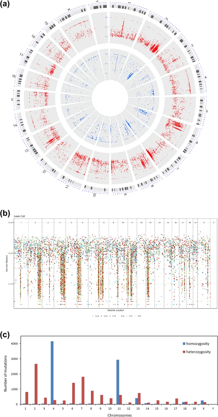
Whole-exome sequencing (WES) revealed that the LLC-derived C57BL mouse represents a previously unrecognized subline closely related to C57BL/6J, which they designated C57BL/6L ("L" for Lewis). Due to the absence of matched normal tissue, somatic mutations were identified by comparing LLC against the C57BL/6J reference genome, capturing variants acquired during tumorigenesis and those accumulated during laboratory passaging since 1951. The analysis identified 18,664 single nucleotide variants (SNVs) and 2,225 insertions/deletions (InDels). Coding regions harbored 5,082 SNVs, with nearly half causing non-synonymous changes (2,201 missense, 63 nonsense). Most InDels (95.6%) were in non-coding regions, while coding InDels predominantly resulted in frameshift or non-frameshift alterations. Mutation distribution was highly uneven, with prominent regional mutation clusters (≥6 consecutive substitutions within ≤10 kb) concentrated on chromosomes 2, 4, 6, 7, 8, 9, 11, and 13 (Fig. 1b), suggesting frequent structural rearrangements. Approximately 38% of all variants were homozygous, clustering primarily on chromosomes 4 and 11 (Fig. 1c). These clusters coincided with regional mutation clusters and likely arose through loss of heterozygosity (LOH) during genomic rearrangements. These findings define the unique mutational architecture of LLC and provide a critical genomic context for its use as a preclinical model.
Ask a Question
Write your own review
Description: C57BL/6-GFP Mouse Skeletal Muscle Microvascular Endothelial Cells from Creative Bioarray are isolated from C57BL/6-Tg (CAG-EGFP) 1Osb/J mouse skeletal muscle tissue of pathogen-free laboratory mice. ...
Description: eNOS KO Mouse Stomach Epithelial Cells from Creative Bioarray are isolated from stomach tissue of pathogen-free laboratory mice. eNOS KO Mouse Stomach Epithelial Cells are grown in a T25 tissue ...
Description: eNOS KO Mouse Liver Fibroblasts from Creative Bioarray are isolated from liver tissue of pathogen-free laboratory mice. eNOS KO Mouse Liver Fibroblasts are grown in T75 tissue culture flasks ...
Description: C57BL/6-GFP Mouse Corneal Epithelial Cells from Creative Bioarray are isolated from C57BL/6-GFP-Tg(CAG-EGFP)1Osb/J mouse corneal tissue of pathogen-free laboratory mice. C57BL/6-GFP Mouse Corneal ...
Description: BALB/c Mouse Retinal Microvascular Endothelial Cells from Creative Bioarray are isolated from retinal tissue of pathogen-free laboratory mice. BALB/c Mouse Retinal Microvascular Endothelial Cells are ...