
C57BL/6 Mouse Tracheal and Bronchial Epithelial Cells
Cat.No.: CSC-C9087J
Species: Mouse
Source: Bronchus; Trachea
Cell Type: Epithelial Cell
- Specification
- Background
- Scientific Data
- Q & A
- Customer Review
Primary C57BL/6 Mouse Tracheal and Bronchial Epithelial Cells obtained from trachea and bronchial tissues of C57BL/6 mice, a prevalent inbred mouse strain used in biomedical research. These cells provide a physiologically realistic in vitro model to research airway biology, respiratory inflammation, epithelial barrier function, and host-pathogen interactions. They often show distinctive epithelial shape such as cobblestone and express airway epithelial markers linked with mucociliary differentiation and innate immune responses.
C57BL/6 mouse airway epithelial cells have been widely employed in respiratory illness models including asthma, chronic obstructive pulmonary disease (COPD), pulmonary fibrosis, and viral infection. These cells may be cultured under air-liquid interface (ALI) conditions and can develop into a functioning airway epithelium with ciliated and mucus generating cell populations. They are useful for research of mucociliary clearance and epithelial remodeling.
These cells are also commonly used in studies of cytokine signaling, oxidative stress, epithelial permeability, and innate immune activation induced by allergens, pollutants, or respiratory pathogens such as influenza virus and SARS-CoV-2. Moreover, C57BL/6 mice Tracheal and Bronchial Epithelial Cells are invaluable for mechanistic investigations in gene regulation, inflammatory signaling pathways, and preclinical treatment evaluation in respiratory research, as they are compatible with genetically modified mice models.
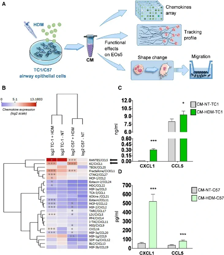
Airway Epithelial Cells Drive Eosinophil Migration via Chemokine Secretion
Eosinophils are a hallmark of allergic asthma, but how eosinophils interact with airway epithelial cells (AECs) before infiltration into tissues is unknown. Raggi et al. set out to delineate the interplay between eosinophils and AECs in IL-33-mediated inflammation.
Using TC1 (a murine lung AT2-derived tumoral cell line) and C57BL/6 Mouse Primary Tracheal and Bronchial Epithelial Cells (hereinafter referred to as C57) stimulated with HDM, they reproduced the asthmatic airway environment in vitro (Fig. 1A). HDM activation resulted in a considerable increase in the secretion of eosinophil-attracting chemokines, such as CXCL1 (3-fold in TC1, 10-fold in C57) and CCL5, compared with untreated cells (Fig. 1B-D).
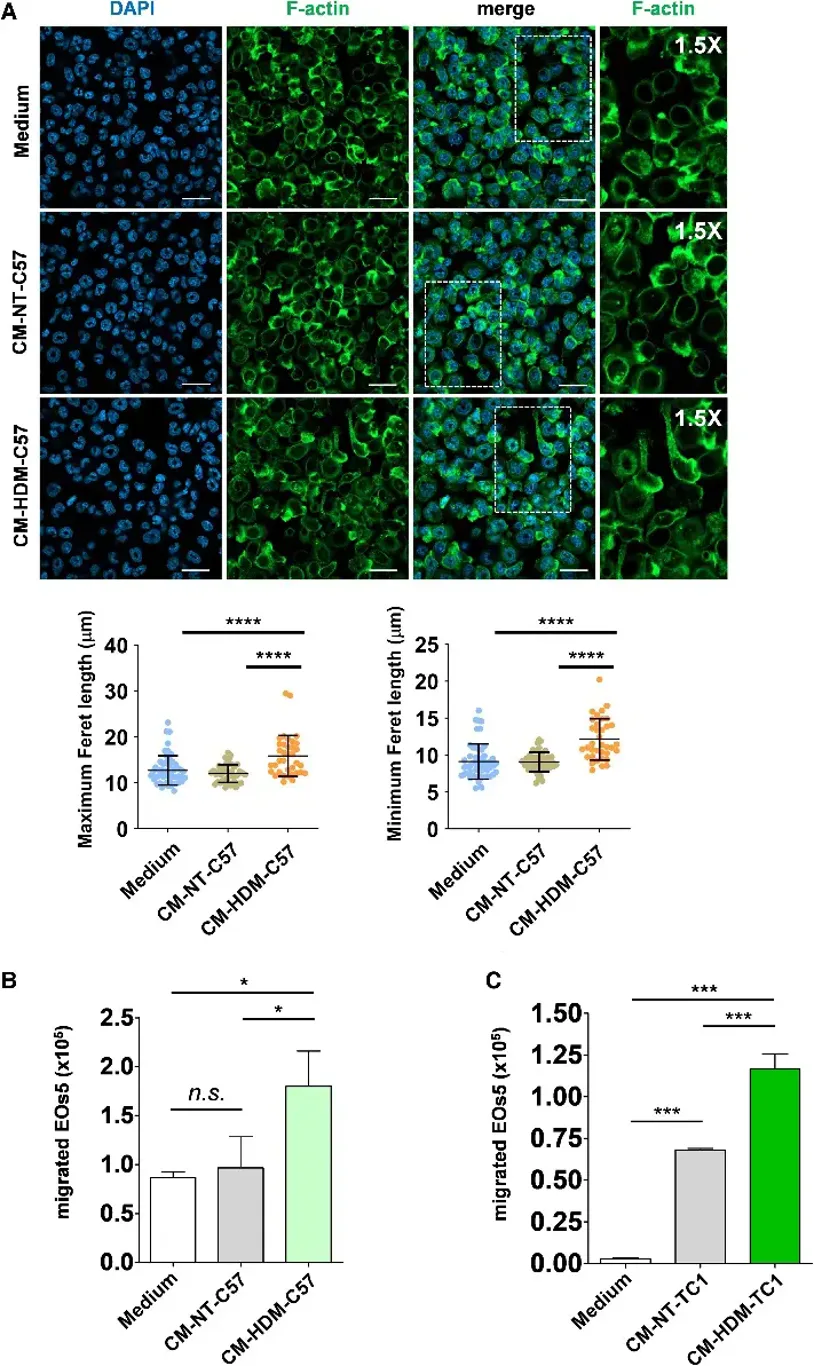
Eos5 cells generated from bone marrow were used to analyze eosinophil migration. Exposure to conditioned medium from HDM-stimulated C57 cells (CM-HDM-C57) elicited morphological alterations associated with migratory activation, i.e. an elongated form and elevated Feret values (Fig. 2A). Eos5 cells showed considerably increased migration toward CM-HDM-C57 compared to controls (Fig. 2B). CM from HDM-stimulated TC1 cells (CM-HDM-TC1) presented similar findings (Fig. 2C). These data collectively suggest that HDM-activated AECs generate a chemotactic gradient that makes eosinophils migratory.
Ask a Question
Write your own review

Description: C57BL/6-GFP Mouse Skeletal Muscle Microvascular Endothelial Cells from Creative Bioarray are isolated from C57BL/6-Tg (CAG-EGFP) 1Osb/J mouse skeletal muscle tissue of pathogen-free laboratory mice. ...

Description: eNOS KO Mouse Stomach Epithelial Cells from Creative Bioarray are isolated from stomach tissue of pathogen-free laboratory mice. eNOS KO Mouse Stomach Epithelial Cells are grown in a T25 tissue ...

Description: eNOS KO Mouse Liver Fibroblasts from Creative Bioarray are isolated from liver tissue of pathogen-free laboratory mice. eNOS KO Mouse Liver Fibroblasts are grown in T75 tissue culture flasks ...
Description: C57BL/6-GFP Mouse Corneal Epithelial Cells from Creative Bioarray are isolated from C57BL/6-GFP-Tg(CAG-EGFP)1Osb/J mouse corneal tissue of pathogen-free laboratory mice. C57BL/6-GFP Mouse Corneal ...

Description: BALB/c Mouse Retinal Microvascular Endothelial Cells from Creative Bioarray are isolated from retinal tissue of pathogen-free laboratory mice. BALB/c Mouse Retinal Microvascular Endothelial Cells are ...