
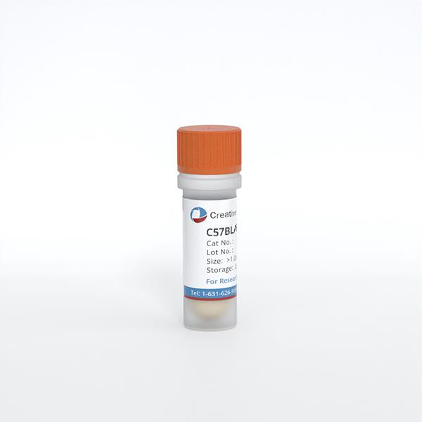
C57BL/6 Mouse Skeletal Muscle Microvascular Endothelial Cells
Cat.No.: CSC-C8336W
Species: Mouse
Source: Skeletal Muscle
Cell Type: Endothelial Cell; Microvascular Cell
- Specification
- Background
- Scientific Data
- Q & A
- Customer Review
C57BL/6 mouse primary skeletal muscle microvascular endothelial cells (MMECs) exhibit the characteristic cobblestone morphology of endothelial monolayers and express canonical endothelial markers including CD31/PECAM-1, VE-cadherin (CD144), and von Willebrand factor (vWF), while remaining negative for hematopoietic marker CD45.
A defining advantage of C57BL/6 MMECs is their retention of in vivo-like functional properties-a feature often lost in immortalized lines. These cells can be expanded for 3-6 passages without phenotypic drift, maintaining their capacity to uptake acetylated low-density lipoprotein (ac-LDL), produce nitric oxide (NO), and form capillary-like vascular tubes in vitro. When transplanted subcutaneously or intramuscularly, they form functional microvessels that integrate with the host vasculature, underscoring their therapeutic angiogenic potential.
C57BL/6 MMECs serve as an indispensable platform for studying endothelial dysfunction in metabolic and vascular diseases, angiogenesis, transendothelial resistance (TER), cell adhesion, and migration. Amenable to genetic modification and available in GFP-expressing or Cas9-expressing derivatives, these cells remain a gold-standard ex vivo model bridging basic microvascular biology and translational therapeutic development.
miRNA-6236 Regulation of Postischemic Skeletal Muscle Angiogenesis
In the present study, Mani, Arul M., et al. (2024) used an unbiased approach by performing miRNA sequencing of RNA isolated from ischemic and nonischemic hind limbs from mice following hind-limb ischemia (HLI). They identified a novel miRNA, miRNA-6236 (miR-6236), whose expression was significantly elevated in the ischemic limbs when compared with nonischemic limbs. Little is known about the role of miR-6236 in general, and its role in postischemic angiogenesis has not been studied. Angiogenesis is a critical component of postischemic recovery following HLI, and endothelial cells play a key role. They therefore investigated the role of miR-6236 in ischemic mouse endothelial cells.
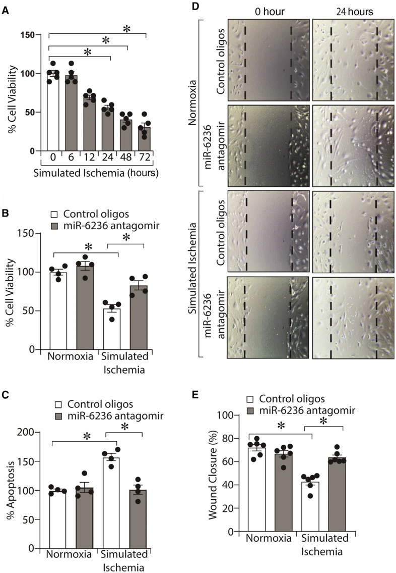
In primary mouse and human endothelial cells, they studied the effect of simulated ischemia on miR-6236 expression and assessed its role in cell viability, apoptosis, migration, and tube formation during ischemia. In vitro simulated ischemia-enhanced miR-6236 expression in mouse and human endothelial cells, whereas its inhibition improved viability, migration, tube formation, and reduced apoptosis. Their results show for the first time that miR-6236 plays a key role in regulating postischemic perfusion recovery and angiogenesis.
Ask a Question
Write your own review

Description: C57BL/6-GFP Mouse Skeletal Muscle Microvascular Endothelial Cells from Creative Bioarray are isolated from C57BL/6-Tg (CAG-EGFP) 1Osb/J mouse skeletal muscle tissue of pathogen-free laboratory mice. ...
Description: eNOS KO Mouse Stomach Epithelial Cells from Creative Bioarray are isolated from stomach tissue of pathogen-free laboratory mice. eNOS KO Mouse Stomach Epithelial Cells are grown in a T25 tissue ...
Description: eNOS KO Mouse Liver Fibroblasts from Creative Bioarray are isolated from liver tissue of pathogen-free laboratory mice. eNOS KO Mouse Liver Fibroblasts are grown in T75 tissue culture flasks ...

Description: C57BL/6-GFP Mouse Corneal Epithelial Cells from Creative Bioarray are isolated from C57BL/6-GFP-Tg(CAG-EGFP)1Osb/J mouse corneal tissue of pathogen-free laboratory mice. C57BL/6-GFP Mouse Corneal ...

Description: BALB/c Mouse Retinal Microvascular Endothelial Cells from Creative Bioarray are isolated from retinal tissue of pathogen-free laboratory mice. BALB/c Mouse Retinal Microvascular Endothelial Cells are ...