
C57BL/6 Mouse Primary Tracheal Epithelial Cells
Cat.No.: CSC-C4248X
Species: Mouse
Source: Trachea
Cell Type: Epithelial Cell
- Specification
- Background
- Scientific Data
- Q & A
- Customer Review
Mouse Primary Tracheal Epithelial Cells can be used in assays of cell to cell adhesion and migration. Standard biochemical procedures performed with epithelial cell cultures include RT-PCR, Western blotting, immunoprecipitation, immunofluorescent staining or immunofluorescent flow cytometry or generating cell derivatives for desired research applications.
C57BL/6 Mouse Primary Tracheal Epithelial Cells are obtained directly from the trachea of the C57BL/6 mouse strain, a genetically well-characterized and commonly used inbred model in biomedical research. These cells make up the mucosal lining of the upper airway and are critical to the maintenance of respiratory homeostasis via mucociliary clearance and barrier protection.
When grown under adherent circumstances, these cells display a distinctive epithelial shape in culture, developing as a polarized monolayer of cobblestone-like cells. They are usually cultivated in a specialized basal medium supplemented with growth factors, hormones and serum to maintain their original phenotype. Routine maintenance is performed in a humidified atmosphere of 37 °C with 5% carbon dioxide. Subculturing is done using a moderate protease solution.
Given their physiological importance these cells are an invaluable model to research airway biology, ciliary function and host-pathogen interactions, in particular in the context of viral infections such as influenza. They are also widely employed in studies of inflammatory responses, toxicological assessment and pathophysiology of chronic respiratory disorders, providing a species-specific platform that supports human bronchial epithelial cell models.
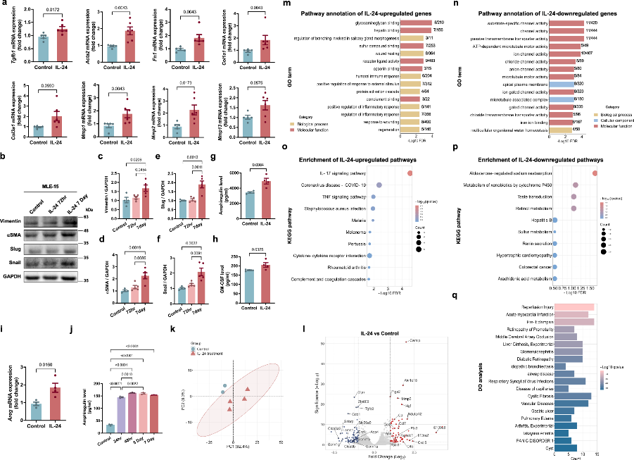
L-24 Specifically Targets Airway Epithelial Cells and Promotes Inflammatory Responses and Remodeling Processes
Asthma is a disease of persistent inflammation and airway remodeling, yet current medications seldom restore structural damage. IL-24 has been implicated in neutrophilic asthma, although its significance in eosinophilic asthma is unknown. Wu's team used IL-24 knockout mice in OVA/HDM models to determine the role of IL-24 to pulmonary disease as a possible therapeutic target.
They developed an ex vivo tracheal epithelial cell culture mimicking in vivo circumstances that provides a biologically relevant platform to study airway disorders. IL-24 stimulation consistently increased amphiregulin production in primary tracheal epithelial cells (Fig. 1i, j) and generated different transcriptome changes as shown by PCA (Fig. 1k). Differential gene expression analysis revealed a total of 138 DEGs, including 61 upregulated and 77 downregulated (Fig. 1l).
Among the upregulated genes were S100a8, C3, Ccl2, Cxcl5, and Mmp3, which are known to enhance airway inflammation and remodeling in asthma (Fig. 1l). However, IL-24 repressed important barrier maintenance genes, including epithelial sodium channels (Scnn1a/b/g), anion transporters (Slc26a9) and Aqp4 (Fig. 1l). These data suggest that IL-24 exacerbates airway disease by concurrently inducing a pro-inflammatory milieu and impairing epithelial barrier integrity.
Ask a Question
Write your own review

Description: C57BL/6-GFP Mouse Skeletal Muscle Microvascular Endothelial Cells from Creative Bioarray are isolated from C57BL/6-Tg (CAG-EGFP) 1Osb/J mouse skeletal muscle tissue of pathogen-free laboratory mice. ...

Description: eNOS KO Mouse Stomach Epithelial Cells from Creative Bioarray are isolated from stomach tissue of pathogen-free laboratory mice. eNOS KO Mouse Stomach Epithelial Cells are grown in a T25 tissue ...

Description: eNOS KO Mouse Liver Fibroblasts from Creative Bioarray are isolated from liver tissue of pathogen-free laboratory mice. eNOS KO Mouse Liver Fibroblasts are grown in T75 tissue culture flasks ...
Description: C57BL/6-GFP Mouse Corneal Epithelial Cells from Creative Bioarray are isolated from C57BL/6-GFP-Tg(CAG-EGFP)1Osb/J mouse corneal tissue of pathogen-free laboratory mice. C57BL/6-GFP Mouse Corneal ...

Description: BALB/c Mouse Retinal Microvascular Endothelial Cells from Creative Bioarray are isolated from retinal tissue of pathogen-free laboratory mice. BALB/c Mouse Retinal Microvascular Endothelial Cells are ...