
C57BL/6 Mouse Primary Proximal Tubular Epithelial Cells
Cat.No.: CSC-C4256X
Species: Mouse
Source: Kidney
Cell Type: Epithelial Cell
- Specification
- Background
- Scientific Data
- Q & A
- Customer Review
Mouse Primary Proximal Tubular Epithelial Cells can be used in assays of cell to cell adhesion and migration. Standard biochemical procedures performed with epithelial cell cultures include RT-PCR, Western blotting, immunoprecipitation, immunofluorescent staining or immunofluorescent flow cytometry or generating cell derivatives for desired research applications.
C57BL/6 Mouse Primary Proximal Tubular Epithelial Cells are isolated from the renal cortex of C57BL/6 inbred mice and represent the portion of nephron responsible for bulk reabsorption of solutes and water. They are polarized epithelial cells with a rich brush border and express characteristic markers such as Lotus tetragonolobus lectin (LTL), Aquaporin-1 (AQP1) and Na+/K+-ATPase.
These cells are cultured in vitro on a collagen or laminin coated substrate, and maintained in a nutrient rich basal medium supplemented with serum, insulin, transferrin and selenium (ITS). The standard culture conditions are 37 °C and 5% CO₂. To preserve phenotype, cultures should be maintained at sub-confluence and passaged sparingly (usually < 5 passages), as dedifferentiation occurs rapidly.
They are mainly used for nephrotoxicity screening and studies of diabetic kidney disease, modelling mechanisms such as albuminuria, oxidative stress and tubular-interstitial fibrosis. They are also important for drug transporter function studies (e.g., OATs, OCTs) and renal drug metabolism. Due to the defined C57BL/6 genetic background, they are ideal for aligning in vitro findings with in vivo murine models of acute kidney injury (AKI) and chronic kidney disease (CKD).
Expression of SGLT2 in the Cultured Tubular Epithelial Cells
While SGLT2 inhibitors are standard diabetes treatments, the mechanisms driving renal SGLT2 overexpression remain unclear. Seki et al. previously linked periodontal pathogens to diabetic nephropathy via TLR signaling. Given that Fusobacterium nucleatumis a potent TLR4 activator in colorectal disease, they investigated whether it exacerbates diabetes by inducing renal SGLT2.
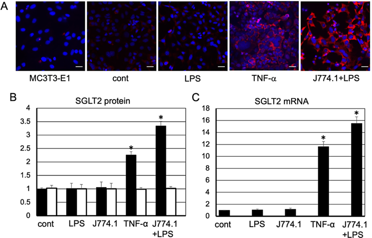
Immunostaining revealed minimal SGLT2 expression in untreated tubular epithelial cells or those exposed to Fn-LPS alone or co-cultured with J774.1 macrophages. However, robust SGLT2 staining was observed in cells treated with TNF-α or co-cultured with J774.1 in the presence of Fn-LPS (Fig. 1). Cell viability remained unaffected across all conditions.
Protein quantification showed no significant increase in SGLT2 levels with Fn-LPS or macrophage co-culture alone compared to controls. In contrast, TNF-α treatment significantly upregulated SGLT2 protein, and this effect was further potentiated by co-culture with Fn-LPS-stimulated J774.1 cells (Fig. 1). Similarly, SGLT2 mRNA levels were significantly elevated by TNF-α alone and were further increased by co-culture with Fn-LPS-stimulated macrophages compared to TNF-α alone (Fig. 1). These findings suggest that F. nucleatum exacerbates hyperglycemia by amplifying TNF-α-mediated SGLT2 expression in renal tubules.
Ask a Question
Write your own review
Description: C57BL/6-GFP Mouse Skeletal Muscle Microvascular Endothelial Cells from Creative Bioarray are isolated from C57BL/6-Tg (CAG-EGFP) 1Osb/J mouse skeletal muscle tissue of pathogen-free laboratory mice. ...
Description: eNOS KO Mouse Stomach Epithelial Cells from Creative Bioarray are isolated from stomach tissue of pathogen-free laboratory mice. eNOS KO Mouse Stomach Epithelial Cells are grown in a T25 tissue ...
Description: eNOS KO Mouse Liver Fibroblasts from Creative Bioarray are isolated from liver tissue of pathogen-free laboratory mice. eNOS KO Mouse Liver Fibroblasts are grown in T75 tissue culture flasks ...
Description: C57BL/6-GFP Mouse Corneal Epithelial Cells from Creative Bioarray are isolated from C57BL/6-GFP-Tg(CAG-EGFP)1Osb/J mouse corneal tissue of pathogen-free laboratory mice. C57BL/6-GFP Mouse Corneal ...
Description: BALB/c Mouse Retinal Microvascular Endothelial Cells from Creative Bioarray are isolated from retinal tissue of pathogen-free laboratory mice. BALB/c Mouse Retinal Microvascular Endothelial Cells are ...