
C57BL/6 Mouse Primary Liver Fibroblasts
Cat.No.: CSC-C1879
Species: Mouse
Source: Liver
Cell Type: Fibroblast
- Specification
- Background
- Scientific Data
- Q & A
- Customer Review
Mouse Primary Liver Fibroblasts are negative for bacteria, yeast, fungi, and mycoplasma. Cells are tested for expression of marker using the antibody of anti-FSP1/S100A4 by immunofluorescence staining. Cells can be expanded for 3-5 passages at a split ratio of 1:2 under the cell culture conditions specified by Creative Bioarray. Repeated freezing and thawing of cells is not recommended.Standard biochemical procedures performed with cell cultures include the assay of cell to cell interaction, RT-PCR, Western blotting, immunoprecipitation, immunofluorescent staining, flow cytometry or generating cell derivatives for desired research applications.
C57BL/6 Mouse Primary Liver Fibroblasts are primary stromal cells derived from the liver tissue of C57BL/6 mice. Liver fibroblasts, as resident mesenchymal cells in the hepatic milieu, participate in the preservation of tissue architecture by producing and organizing extracellular matrix (ECM) components. In addition to their structural role, these cells communicate with hepatocytes, endothelial cells, immune cells, and hepatic stellate cells to regulate tissue homeostasis and repair processes.
Under healthy conditions, liver fibroblasts contribute to the integrity of the hepatic stromal compartment and to matrix turnover. In response to tissue injury, inflammation and metabolic stress, these cells can be triggered and switch their secretory profile to influence extracellular matrix remodeling and local cellular responses. Liver fibroblasts have been established in studies to be a major component in the hepatic microenvironment participating in signaling networks related to fibrosis, wound healing and inflammatory control.
C57BL/6 Mouse Primary Liver Fibroblasts are an in vitro model to study liver stromal biology and cell-matrix interactions. They are frequently used in studies of hepatic fibrosis, extracellular matrix deposition, inflammatory signaling, tissue remodeling and liver regeneration. These cells are also well suited for assessing the impact of cytokines, growth factors and medicinal substances on fibroblast activation and stromal responses.
Biosafety Evaluation of a Graphene Oxide-Captopril Sustained-Release System
Captopril (CAPT), a widely prescribed antihypertensive, faces limitations of rapid clearance and poor aqueous solubility. Graphene oxide (GO) was explored as a nanocarrier to enable sustained CAPT delivery, but its biosafety-particularly at high concentrations and large lateral sizes-requires rigorous validation. Liu's team assessed the cytotoxicity of a GO-CAPT delivery system across three biological dimensions: cell proliferation, structural integrity, and cell death modality, using human colorectal adenocarcinoma (Caco-2) cells and primary mouse hepatic fibroblasts.
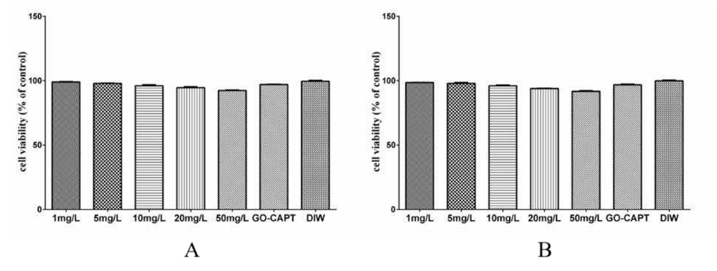
Proliferation assays using the WST-1 kit showed that even at a high GO concentration of 50 mg/L, the cell survival rate in Caco-2 cells remained 92.38%, compared to 99.58% in the deionized water (DIW) control, representing only a 7.2% reduction (Fig. 1A). The GO-CAPT formulation showed a minimal 2.59% reduction versus control (96.99% viability) and displayed no statistically significant impact. Identical trends were observed in primary mouse liver fibroblasts (Fig. 1B), confirming negligible effects on cell proliferation.
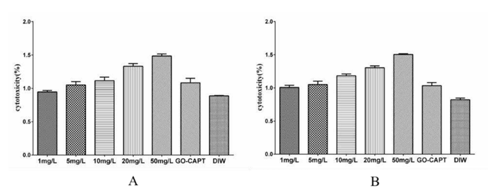
To evaluate structural integrity, lactate dehydrogenase (LDH) release was measured. In Caco-2 cells, cytotoxicity in the DIW control was 0.89%, while GO at 50 mg/L induced only 1.48% cytotoxicity-a 0.59% difference (Fig. 2A). The GO-CAPT group exhibited 1.08% cytotoxicity, differing by just 0.19% from the control. Again, primary fibroblasts mirrored this low-impact profile (Fig. 2B). These results demonstrate that GO with a large lateral size (1-2 µm) and concentrations up to 50 mg/L does not significantly impair cell proliferation or membrane integrity. Furthermore, the GO-CAPT delivery system exhibits excellent cytocompatibility, supporting its potential as a safe and effective platform for sustained captopril release in biomedical applications.
Ask a Question
Write your own review


Description: C57BL/6-GFP Mouse Skeletal Muscle Microvascular Endothelial Cells from Creative Bioarray are isolated from C57BL/6-Tg (CAG-EGFP) 1Osb/J mouse skeletal muscle tissue of pathogen-free laboratory mice. ...

Description: eNOS KO Mouse Stomach Epithelial Cells from Creative Bioarray are isolated from stomach tissue of pathogen-free laboratory mice. eNOS KO Mouse Stomach Epithelial Cells are grown in a T25 tissue ...

Description: eNOS KO Mouse Liver Fibroblasts from Creative Bioarray are isolated from liver tissue of pathogen-free laboratory mice. eNOS KO Mouse Liver Fibroblasts are grown in T75 tissue culture flasks ...
Description: C57BL/6-GFP Mouse Corneal Epithelial Cells from Creative Bioarray are isolated from C57BL/6-GFP-Tg(CAG-EGFP)1Osb/J mouse corneal tissue of pathogen-free laboratory mice. C57BL/6-GFP Mouse Corneal ...

Description: BALB/c Mouse Retinal Microvascular Endothelial Cells from Creative Bioarray are isolated from retinal tissue of pathogen-free laboratory mice. BALB/c Mouse Retinal Microvascular Endothelial Cells are ...