
C57BL/6 Mouse Pancreatic Microvascular Endothelial Cells
Cat.No.: CSC-C8330W
Species: Mouse
Source: Pancreas
Cell Type: Endothelial Cell; Microvascular Cell
- Specification
- Background
- Scientific Data
- Q & A
- Customer Review
C57BL/6 Mouse Pancreatic Microvascular Endothelial Cells are obtained from the pancreatic tissue microvasculature from C57BL/6 mice. These cells compose the inner layer of pancreatic micro-vessels and are vital for maintaining vascular homeostasis, controlling the flow of nutrients and oxygen and facilitating communication between the vascular system and surrounding pancreatic cells. Microvascular endothelial cells are important for the structural and functional integrity of the pancreatic microenvironment of the pancreas. They are involved in regulation of vascular permeability, inflammation, angiogenesis and tissue remodeling. Moreover, endothelial cell-pancreatic islet cell interactions are known to be critical in appropriate endocrine function and metabolic regulation.
C57BL/6 Mouse Pancreatic Microvascular Endothelial Cells are useful in vitro models to research pancreatic vascular biology and endothelial cellular behavior under healthy and pathological settings. These cells have been used in studies on diabetes, islet vascularization, endothelial dysfunction, inflammation, oxidative stress and pancreatic tissue damage. They are also well suited for studying endothelial responses to cytokines, growth factors, metabolic stress, and pharmaceutical substances.
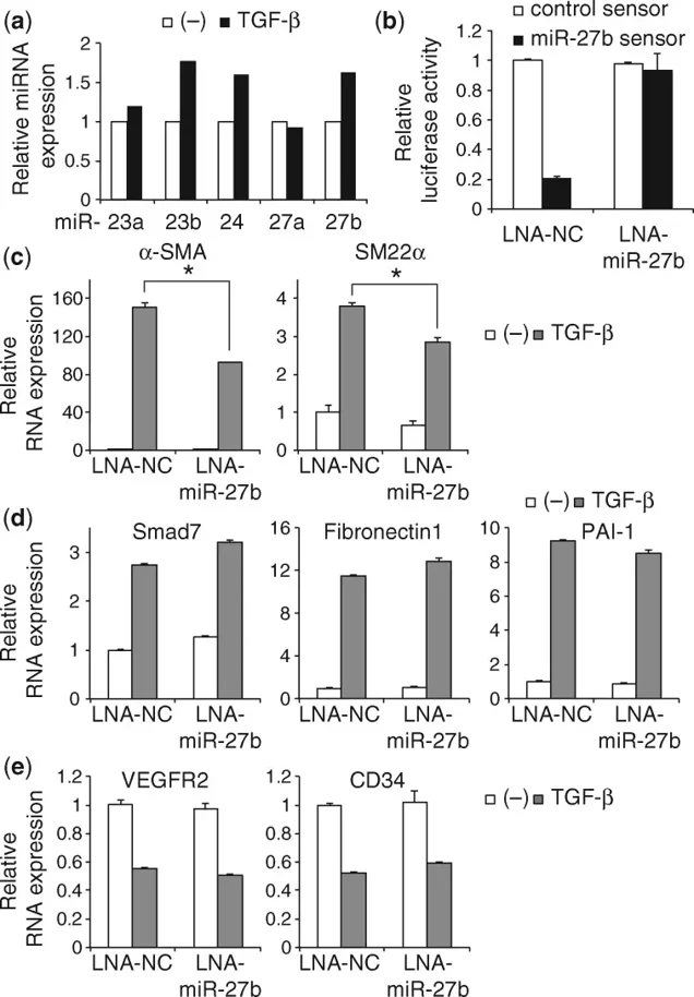
miR-27b Promotes TGF-β-Induced Endothelial-Mesenchymal Transition in Pancreatic Endothelial Cells
MicroRNAs (miRNAs) are key regulators of epithelial-mesenchymal transition (EMT) and endothelial-mesenchymal transition (EndMT). In this study, miRNA microarray analysis identified that transforming growth factor-β (TGF-β) significantly upregulated miR-23b, miR-24, and miR-27b in MS-1 mouse pancreatic microvascular endothelial cells, while effects on miR-23a, miR-24-2, and miR-27a were negligible (Fig. 1a).
To determine the role of miR-27b in EndMT, they inhibited endogenous miR-27b using a locked nucleic acid (LNA) inhibitor. LNA-miR-27b effectively abolished miR-27b activity in MS-1 cells (Fig. 1b) and suppressed TGF-β-induced expression of mesenchymal markers α-SMA and SM22α (Fig. 1c). In contrast, miR-27b inhibition did not attenuate TGF-β induction of canonical Smad target genes (Smad7, Fibronectin1, PAI-1) or prevent downregulation of endothelial markers (VEGFR2, CD34).
These findings demonstrate that TGF-β selectively upregulates the miR-23b/24-1/27b cluster in MS-1 cells and that miR-27b specifically enhances mesenchymal gene expression during EndMT, independent of canonical Smad signaling. This identifies miR-27b as a positive regulator of TGF-β-driven EndMT in pancreatic endothelial cells.
Ask a Question
Write your own review
Description: C57BL/6-GFP Mouse Skeletal Muscle Microvascular Endothelial Cells from Creative Bioarray are isolated from C57BL/6-Tg (CAG-EGFP) 1Osb/J mouse skeletal muscle tissue of pathogen-free laboratory mice. ...

Description: eNOS KO Mouse Stomach Epithelial Cells from Creative Bioarray are isolated from stomach tissue of pathogen-free laboratory mice. eNOS KO Mouse Stomach Epithelial Cells are grown in a T25 tissue ...

Description: eNOS KO Mouse Liver Fibroblasts from Creative Bioarray are isolated from liver tissue of pathogen-free laboratory mice. eNOS KO Mouse Liver Fibroblasts are grown in T75 tissue culture flasks ...

Description: C57BL/6-GFP Mouse Corneal Epithelial Cells from Creative Bioarray are isolated from C57BL/6-GFP-Tg(CAG-EGFP)1Osb/J mouse corneal tissue of pathogen-free laboratory mice. C57BL/6-GFP Mouse Corneal ...

Description: BALB/c Mouse Retinal Microvascular Endothelial Cells from Creative Bioarray are isolated from retinal tissue of pathogen-free laboratory mice. BALB/c Mouse Retinal Microvascular Endothelial Cells are ...