
Immortalized Human Mammary Epithelial Cells-GFP (MCF-12A)
Cat.No.: CSC-I2277Z
Species: homo sapiens
Morphology: Polygonal
Culture Properties: Adherent
- Specification
- Background
- Scientific Data
- Q & A
- Customer Review
Immortalized Human Mammary Epithelial Cells‑GFP (MCF‑12A‑GFP) is a non‑tumorigenic, spontaneously immortalized breast‑epithelial cell line engineered for expression of enhanced green fluorescent protein for live‑cell imaging. Parental MCF‑12A cells were obtained from reduction mammoplasty tissue from a white, nulliparous woman with fibrocystic breast disease that contained focal intraductal hyperplasia. They were established by long‑term culture in serum‑free, low‑calcium medium, a method of immortalization that maintains an adherent, epithelial morphology.
GFP‑tagged derivatives are usually produced by lentiviral or plasmid transduction of a GFP cassette followed by antibiotic selection (puromycin or G418) to select for stable, brightly fluorescent clones. GFP expression allows for real‑time monitoring of cell proliferation, migration and three‑dimensional morphogenesis, and allows for co‑culture studies in which normal mammary epithelium can be distinguished from malignant counterparts. Functionally, MCF‑12A cells are estrogen‑receptor negative, express a combination of epithelial (CK8, CK14, E‑cadherin) and mesenchymal markers, and form lumenized acini or duct‑like structures in collagen or Matrigel matrices. MCF‑12A cells are widely used as a "normal" control in breast‑cancer research, in studies of ETS transcription‑factor‑mediated transformation and in radiobiology studies of low‑energy X‑rays.
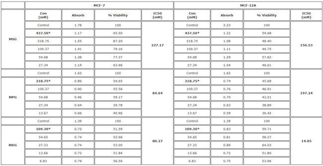
Effects of Some Flavor Enhancer Food Additives on Expression of Cancer-Related Genes in MCF-7 and MCF-12A Cells
Food additives are essential for food preservation and enhancement. Here, Bolukbasi et al. treated MCF-7 and MCF-12A cells with various concentrations of MSG, MPG, and MDG for 24 hours to assess cytotoxicity and gene expression changes. The 50% inhibitory concentration (IC50) value was calculated by determining the MTT results and the applied dose and % cell viability curve with the Microsoft Excel (Cytotoxicity = test absorbance value/control absorbance value average x100). The experiments were repeated three times for each chemical concentration and the results are shown in Figure 1. According to the results, IC50 values of MSG, MPG and MDG were measured as 227.17, 84.64 and 80.27 mM for MCF-7 and 156.53, 197.24 and 14.05 mM for MCF-12A, respectively. The results showed that all three food additives have the ability to induce cytotoxicity in MCF-7 and MCF-12A cell lines (Fig. 1).
Ask a Question
Write your own review
Description: Nasal epithelial cells form the outermost protective layer against environmental factors. They clean, humidify, and warm inhaled air. They produces mucus, which binds particles that are subsequently ...

Description: Immortalized Human Corneal Epithelial Cells-SV40 have been obtained immortalizing Human Corneal Epithelial Cells with Lenti-SV40 Lentivirus. Immortalized cells were controlled passaging side by side ...
Description: Immortalized Human Lymphatic Endothelial Cells-SV40 were developed from human tissues transduced with a lentiviral expression vector containing the SV40T gene. The cell line was continuously cultured ...
Description: Immortalized Human Retinal Pigment Epithelial Cells were isolated from neonatal human globes and spontaneously immortalized in culture bypassing crisis. They retained typical morphology of the ...
- Adipose Tissue-Derived Stem Cells
- Human Neurons
- Mouse Probe
- Whole Chromosome Painting Probes
- Hepatic Cells
- Renal Cells
- In Vitro ADME Kits
- Tissue Microarray
- Tissue Blocks
- Tissue Sections
- FFPE Cell Pellet
- Probe
- Centromere Probes
- Telomere Probes
- Satellite Enumeration Probes
- Subtelomere Specific Probes
- Bacterial Probes
- ISH/FISH Probes
- Exosome Isolation Kit
- Human Adult Stem Cells
- Mouse Stem Cells
- iPSCs
- Mouse Embryonic Stem Cells
- iPSC Differentiation Kits
- Mesenchymal Stem Cells
- Immortalized Human Cells
- Immortalized Murine Cells
- Cell Immortalization Kit
- Adipose Cells
- Cardiac Cells
- Dermal Cells
- Epidermal Cells
- Peripheral Blood Mononuclear Cells
- Umbilical Cord Cells
- Monkey Primary Cells
- Mouse Primary Cells
- Breast Tumor Cells
- Colorectal Tumor Cells
- Esophageal Tumor Cells
- Lung Tumor Cells
- Leukemia/Lymphoma/Myeloma Cells
- Ovarian Tumor Cells
- Pancreatic Tumor Cells
- Mouse Tumor Cells