
Immortalized Human Osteoblasts-SV40
Cat.No.: CSC-I9173L
Species: Homo sapiens
Source: Femoral Bone tissue
Morphology: Multipolar
Culture Properties: Adherent
- Specification
- Background
- Scientific Data
- Q & A
- Customer Review
Note: Never can cells be kept at -20 °C.
CIK-HT003 HT® Lenti-SV40T Immortalization Kit
Immortalized Human Osteoblasts-SV40 immortalized cell lines are osteoblastic cells transformed with the SV40 large T antigen. The SV40 large T antigen knocks out certain cell cycle proteins, including p53 and Rb which have increased apoptosis during cell division, allowing for cellular immortality.
Immortalized osteoblasts have osteoblastic cell morphology and express markers such as alkaline phosphatase (ALP), osteocalcin, and type I collagen. They also retain the ability to differentiate and mineralize when given osteogenic media. Similar to primary osteoblasts, SV40 osteoblasts are able to respond to vitamin D and parathyroid hormone treatment. Immortalized human osteoblasts have been utilized in many models including development, bone remodeling, and bone diseases such as osteoporosis, bone tumor, and metabolic bone disease. These cells are also used as a control model for osteosarcoma. Immortalized human osteoblast cells have been frequently used in the biomaterials and tissue engineering industry to test for biocompatibility of scaffolds and implantable materials along with cell attachment osteoinductivity. Due to their stability these cells have been used in drug discovery research to identify inhibitors or promoters of bone formation and mineralization.
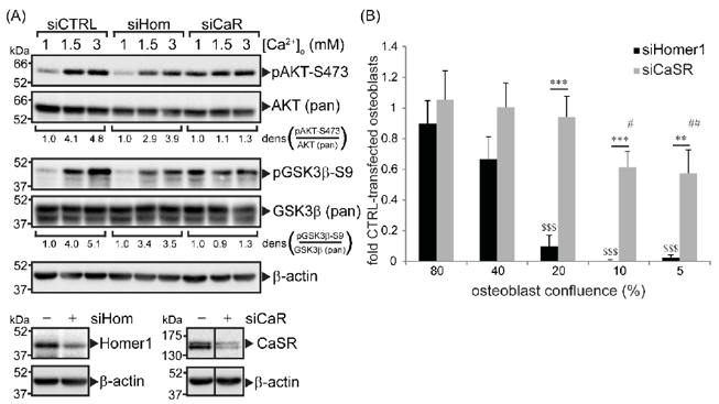
CaSR and Homer1 Were Required for Extracellular Ca2+-Dependent AKT and GSK3β Phosphorylation in Immortalized Primary Human Osteoblasts and Promoted Osteoblast Viability
In human osteoblasts, Homer1 binds to the Calcium-sensing receptor (CaSR) and promotes AKT activation via mTORC2, stabilizing β-catenin and activating mTORC1. Rybchyn et al. further explores the functional relationship between Homer1 and CaSR in primary human osteoblasts.
Using siRNA knockdown in immortalized primary human osteoblasts, we found that depleting either Homer1 or CaSR markedly suppressed extracellular Ca2+-induced phosphorylation of AKT-Ser473 and GSK3β-Ser9, consistent with prior reports (Fig. 1A).
Because AKT is critical for cell survival, they tested the effects of silencing Homer1 or CaSR on osteoblast viability under low-density stress. Both knockdowns reduced cell viability, with Homer1 depletion causing a more pronounced loss. Notably, at seeding densities of 10% or lower, Homer1 silencing completely abolished cell viability, indicating its essential role in osteoblast survival under stress (Fig. 1B).
Ask a Question
Write your own review

Description: Nasal epithelial cells form the outermost protective layer against environmental factors. They clean, humidify, and warm inhaled air. They produces mucus, which binds particles that are subsequently ...
Description: Immortalized Human Splenic Endothelial Cells-SV40 have been obtained immortalizing Human Splenic Endothelial Cells with SV40LT expressing lentiviral particles. Immortalized cells were controlled ...
Description: Immortalized Human Corneal Epithelial Cells-SV40 have been obtained immortalizing Human Corneal Epithelial Cells with Lenti-SV40 Lentivirus. Immortalized cells were controlled passaging side by side ...
Description: Immortalized Human Lymphatic Endothelial Cells-SV40 were developed from human tissues transduced with a lentiviral expression vector containing the SV40T gene. The cell line was continuously cultured ...
Description: Immortalized Human Retinal Pigment Epithelial Cells were isolated from neonatal human globes and spontaneously immortalized in culture bypassing crisis. They retained typical morphology of the ...
- Adipose Tissue-Derived Stem Cells
- Human Neurons
- Mouse Probe
- Whole Chromosome Painting Probes
- Hepatic Cells
- Renal Cells
- In Vitro ADME Kits
- Tissue Microarray
- Tissue Blocks
- Tissue Sections
- FFPE Cell Pellet
- Probe
- Centromere Probes
- Telomere Probes
- Satellite Enumeration Probes
- Subtelomere Specific Probes
- Bacterial Probes
- ISH/FISH Probes
- Exosome Isolation Kit
- Human Adult Stem Cells
- Mouse Stem Cells
- iPSCs
- Mouse Embryonic Stem Cells
- iPSC Differentiation Kits
- Mesenchymal Stem Cells
- Immortalized Human Cells
- Immortalized Murine Cells
- Cell Immortalization Kit
- Adipose Cells
- Cardiac Cells
- Dermal Cells
- Epidermal Cells
- Peripheral Blood Mononuclear Cells
- Umbilical Cord Cells
- Monkey Primary Cells
- Mouse Primary Cells
- Breast Tumor Cells
- Colorectal Tumor Cells
- Esophageal Tumor Cells
- Lung Tumor Cells
- Leukemia/Lymphoma/Myeloma Cells
- Ovarian Tumor Cells
- Pancreatic Tumor Cells
- Mouse Tumor Cells