

Immortalized Human Nasal Epithelial Cells-SV40
Cat.No.: CSC-I9157L
Species: homo sapiens
Source: Nasal Mucosa
Morphology: Polygonal
Culture Properties: Adherent
- Specification
- Background
- Scientific Data
- Q & A
- Customer Review
- Documents
Note: Never can cells be kept at -20 °C.
CIK-HT003 HT® Lenti-SV40T Immortalization Kit
Immortalized Human Nasal Epithelial Cells-SV40 are a nasal epithelial cell line representing an in vitro model of primary human nasal epithelial cells that have been immortalized using SV40 large T antigen. Immortalized Human Nasal Epithelial Cells-SV40 cells will grow as an epithelial monolayer with cobblestone morphology when maintained in standard tissue culture conditions. Under these conditions, the cells will express cytokeratins, E-cadherin, and tight junction proteins (such as ZO-1 and occludin), characteristic of an epithelial barrier. Additionally, when polarized, nasal epithelial cells can perform many barrier functions specific to the upper airway such as production of mucus, innate immune signaling, and secretion of cytokines. Research has previously demonstrated that airway epithelial cell models maintain responsiveness to outside factors such as allergens, pathogens and inflammatory mediators via innate inflammatory signaling pathways (i.e., NF-κB and interferon signaling). Due to this, nasal epithelial cells serve as useful models for disease phenotypes.
Immortalized Human Nasal Epithelial Cells-SV40 cells are commonly used to study respiratory diseases such as allergic rhinitis, chronic rhinosinusitis, and viral infection of the nasal epithelium. Nasal epithelial cells serve as useful models to study host-pathogen interactions, barrier integrity of the nasal epithelium and transport / delivery of pharmaceuticals across the nasal mucosa. Compared to primary nasal epithelial cells, Immortalized Human Nasal Epithelial Cells-SV40 offer increased stability, reproducibility and scalability.
MSC-sEV Restored Nasal Barrier Function in Allergic Rhinitis Via Mir-143-GSK3B in Human Nasal Epithelial Cells
The nasal epithelial barrier is critical for defending against pathogens and allergens, with dysfunction implicated in allergic rhinitis (AR). Mesenchymal stem cell-derived small extracellular vesicles (MSC-sEV) show anti-inflammatory and regenerative potential, but their effects on nasal epithelial cells are unknown. Xu's team investigated MSC-sEV's therapeutic effects on AR and underlying mechanisms.
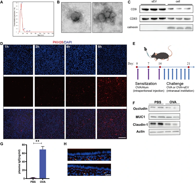
An ovalbumin-induced mouse AR model and immortalized human nasal epithelial cells (HNEpC) were used to assess MSC-sEV effects. Characterization and nanoparticle tracing analysis revealed that MSC-sEVs had an average diameter of 125 nm, with a size distribution between 87 and 200 nm (Fig 1A). Transmission electron microscopy confirmed their spherical morphology (Fig 1B). Western blotting showed enrichment of classical exosomal markers CD63 and CD9, and absence of the negative marker calnexin, compared to cell lysate (Fig 1C). To assess uptake by nasal epithelial cells, PKH26-labeled MSC-sEVs were cocultured with HNEpCs. Increasing PKH26 fluorescence intensity over time confirmed active uptake (Fig 1D). An experimental allergic rhinitis (AR) model was established via OVA intraperitoneal injection and nasal drops (Fig 1E). Western blot analysis showed reduced tight junction proteins (occludin, claudin-1) and mucoprotein MUC1, indicating barrier disruption, while elevated plasma IgE confirmed successful allergic sensitization (Fig 1F, G). In vivo, PKH26-labeled MSC-sEVs delivered intranasally produced positive fluorescence signals in nasal mucosa (Fig 1H). Collectively, they characterized MSC-sEVs, established an AR mouse model, and confirmed active MSC-sEV uptake by epithelial cells both in vitro and in vivo.
Ask a Question
Write your own review
 Immortalized Human Nasal Epithelial Cells-SV40
Description: Immortalized Human Splenic Endothelial Cells-SV40 have been obtained immortalizing Human Splenic Endothelial Cells with SV40LT expressing lentiviral particles. Immortalized cells were controlled ...
Description: Immortalized Human Corneal Epithelial Cells-SV40 have been obtained immortalizing Human Corneal Epithelial Cells with Lenti-SV40 Lentivirus. Immortalized cells were controlled passaging side by side ...

Description: Immortalized Human Lymphatic Endothelial Cells-SV40 were developed from human tissues transduced with a lentiviral expression vector containing the SV40T gene. The cell line was continuously cultured ...
Description: Immortalized Human Retinal Pigment Epithelial Cells were isolated from neonatal human globes and spontaneously immortalized in culture bypassing crisis. They retained typical morphology of the ...
- Adipose Tissue-Derived Stem Cells
- Human Neurons
- Mouse Probe
- Whole Chromosome Painting Probes
- Hepatic Cells
- Renal Cells
- In Vitro ADME Kits
- Tissue Microarray
- Tissue Blocks
- Tissue Sections
- FFPE Cell Pellet
- Probe
- Centromere Probes
- Telomere Probes
- Satellite Enumeration Probes
- Subtelomere Specific Probes
- Bacterial Probes
- ISH/FISH Probes
- Exosome Isolation Kit
- Human Adult Stem Cells
- Mouse Stem Cells
- iPSCs
- Mouse Embryonic Stem Cells
- iPSC Differentiation Kits
- Mesenchymal Stem Cells
- Immortalized Human Cells
- Immortalized Murine Cells
- Cell Immortalization Kit
- Adipose Cells
- Cardiac Cells
- Dermal Cells
- Epidermal Cells
- Peripheral Blood Mononuclear Cells
- Umbilical Cord Cells
- Monkey Primary Cells
- Mouse Primary Cells
- Breast Tumor Cells
- Colorectal Tumor Cells
- Esophageal Tumor Cells
- Lung Tumor Cells
- Leukemia/Lymphoma/Myeloma Cells
- Ovarian Tumor Cells
- Pancreatic Tumor Cells
- Mouse Tumor Cells