
TXM13
Cat.No.: CSC-C9741L
Species: Homo sapiens (Human)
Source: Brain Metastasis
Culture Properties: monolayer
- Specification
- Background
- Scientific Data
- Q & A
- Customer Review
vWA: 16,17
FGA: 22.2,25
Amelogenin: X
TH01: 8
TPOX: 10,11
CSF1P0: 11
D5S818: 12
D13S317: 8,12
D7S820: 8
Shipping Condition: Room Temperature
The TXM-13 (also called TXM13) is a human melanotic melanoma cell line, which was established from a brain metastatic lesion of a melanoma patient. It is a highly tumorigenic and metastatic model, which is widely used in cancer studies. TXM-13 is characterized by its high rate of stable melanin pigmentation. TXM-13 can be maintained in pigmented state with a high efficiency unlike other pigmented melanoma lines (e.g. G361 or SK-MEL-28) that are prone to spontaneous depigmentation during long-term culturing. This stability is explained by the consistent copy number of the tyrosinase gene locus (Chr 11q21). Morphological observation revealed that the cells had a heterogeneous mixture of pigmented and non-pigmented adherent growth. They are usually cultured in a nutrient-rich basal medium supplemented with serum under conventional conditions (37°C, 5% CO2).
TXM-13 displays a stable melanogenic activity and can be used as a tool for researching tyrosinase-mediated melanogenesis, screening depigmenting/whitening drugs, and investigating the pathophysiology of melanoma metastasis, especially to brain.
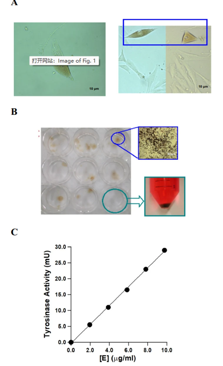
Tyrosinase-Mediated TXM13 Pigmentation During Long-Term Culture
Melanoma exhibits heterogeneous melanin production, yet its molecular determinants remain unclear. Yin's team characterized TXM13 cells, a melanotic melanoma line that displays heterogeneous pigmentation during subculture (Fig. 1A). To isolate highly pigmented cells, they performed single-cell cloning. Cloned cells maintained robust pigmentation upon scale-up (Fig. 1B) and were harvested for analysis.
Tyrosinase activity assays confirmed that the cloned TXM13 cells exhibited specific DOPA oxidase activity in a concentration-dependent manner (Fig. 1C). This demonstrates that the high pigmentation phenotype is directly associated with functional tyrosinase expression, establishing this clonal line as a model for studying melanin regulation.
Ask a Question
Write your own review
- You May Also Need

Description: Established from the primary tumor (right cervical) of a 64-year-old woman with cutaneous melanoma

Description: Established from the lymph node metastasis of a malignant melanoma from a 42-year-old Caucasian woman
Description: Species: human - male, 31 years old, CaucasianIsoenzyme: G6PD, BProduction: melaninHistopathology: melanoma
Description: Epstein-Barr virus-positive cell line established from peripheral blood in 1977 from male patient with melanoma

Description: Established from the primary tumor of a 58-year-old woman with melanoma in 1977

Description: Established from the primary (achromic) cutaneous tumor (left thigh) of a 26-year-old man with malignant melanoma (primary tumor histology: SSM level IV); same patient as cell line IGR-37; described ...
- Adipose Tissue-Derived Stem Cells
- Human Neurons
- Mouse Probe
- Whole Chromosome Painting Probes
- Hepatic Cells
- Renal Cells
- In Vitro ADME Kits
- Tissue Microarray
- Tissue Blocks
- Tissue Sections
- FFPE Cell Pellet
- Probe
- Centromere Probes
- Telomere Probes
- Satellite Enumeration Probes
- Subtelomere Specific Probes
- Bacterial Probes
- ISH/FISH Probes
- Exosome Isolation Kit
- Human Adult Stem Cells
- Mouse Stem Cells
- iPSCs
- Mouse Embryonic Stem Cells
- iPSC Differentiation Kits
- Mesenchymal Stem Cells
- Immortalized Human Cells
- Immortalized Murine Cells
- Cell Immortalization Kit
- Adipose Cells
- Cardiac Cells
- Dermal Cells
- Epidermal Cells
- Peripheral Blood Mononuclear Cells
- Umbilical Cord Cells
- Monkey Primary Cells
- Mouse Primary Cells
- Breast Tumor Cells
- Colorectal Tumor Cells
- Esophageal Tumor Cells
- Lung Tumor Cells
- Leukemia/Lymphoma/Myeloma Cells
- Ovarian Tumor Cells
- Pancreatic Tumor Cells
- Mouse Tumor Cells