
SK-MEL-3
Cat.No.: CSC-C0099
Species: Homo sapiens (Human)
Source: Lymph Node Metastasis
Morphology: adherent epithelial cells growing as monolayer
Culture Properties: monolayer
- Specification
- Background
- Scientific Data
- Q & A
- Customer Review
Immunology: cytokeratin -, desmin -, endothel -, EpCAM -, GFAP -, HMB-45 +, neurofilament -, vimentin +
Viruses: ELISA: reverse transcriptase negative; PCR: EBV -, HBV -, HCV
SK-MEL-3 is a human malignant melanoma cell line, derived from a lymph node metastasis of a patient with cutaneous melanoma. It is one of the related lines in the famous SK-MEL cell series. The cells are in the late stage of melanoma and still have a lot of phenotypic and molecular traits similar to metastatic disease.
At the molecular and immunophenotypic level, SK-MEL-3 cells express the melanoma-associated antigens MART-1 (Melan-A), gp100 (PMEL), and tyrosinase and have been used widely in immunologic studies of melanoma. At the genetic level, this cell line also shares a number of alterations with melanoma in general, such as aberrant MAPK pathway activation, leading to increased proliferation and survival of these cells. Functionally, SK-MEL-3 cells show malignant phenotypes, such as high proliferative capacity and invasive growth. Expression of the tumor-associated antigens MART-1, gp100, and tyrosinase in these cells has made this cell line particularly useful in studies of tumor immunogenicity, tumor antigen presentation, and immune-mediated killing of tumor cells. The SK-MEL-3 cell line is commonly used to study immune checkpoint signaling, cancer vaccines, and adoptive cell therapies as well as for preclinical testing of targeted and immunotherapeutic drugs.
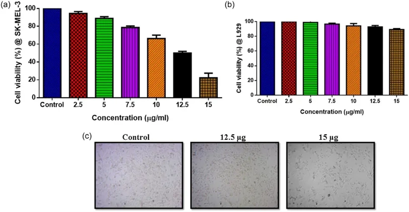
Anticancer Activity of CuO/TiO2-chitosan-farnesol NCs
This investigation synthesizing copper oxide (CuO)-titanium oxide (TiO2)-chitosan-farnesol nanocomposites with potential antibacterial, antifungal, and anticancer properties against Melanoma cells (melanoma cells [SK-MEL-3]).
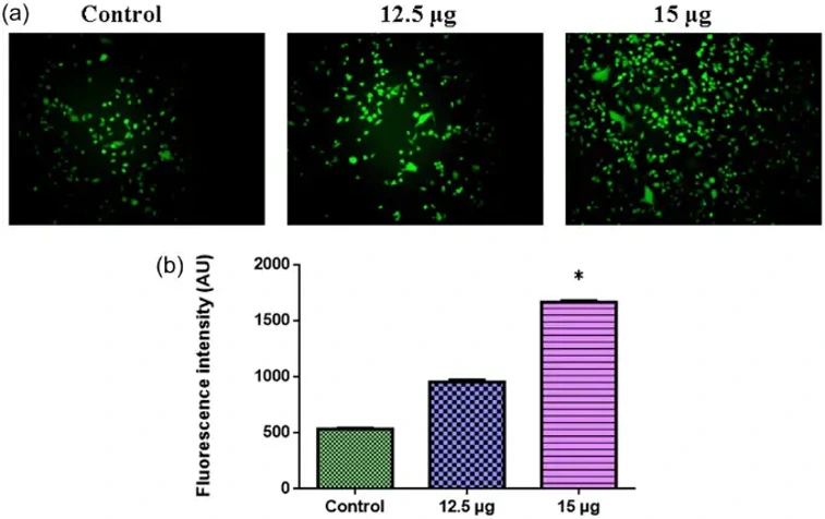
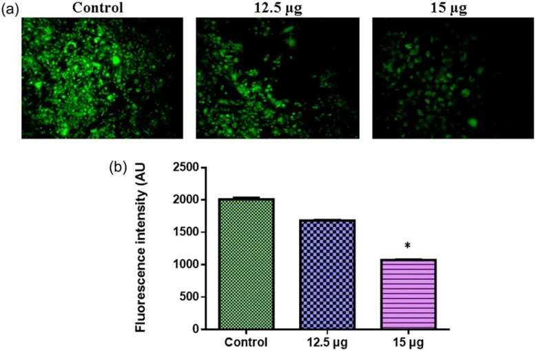
SK-MEL-3 cells were first treated with CuO/TiO2-chitosan-farnesol nanocomposites using MTT assays to determine cytotoxic effect. The IC50 was determined to be 10 and 12.5 µg/mL. IC50 values were used in all subsequent experiments to evaluate the anticancer efficacy (Fig. 1a and b). SK-MEL-3 cells were then treated with CuO/TiO2-chitosan-farnesol nanocomposites at 12.5 and 15 µg/mL for 24 h. Cells were then fixed, stained, and visualized with a fluorescence microscope. Treated cells had significantly higher AO/ETBR staining than control cells (p < 0.05) indicating greater cell damage (Fig. 2a and b). SK-MEL-3 cells were treated with CuO/TiO2-chitosan-farnesol nanocomposites (12.5 and 15 µg/mL) in the dark for 24 hours. ROS production was significantly higher in treated cells between 6 and 12 hours (p < 0.05), while control cells produced little to no ROS. ROS analysis was done three times, with the average data shown in a bar graph (Fig. 3a and b).
Ask a Question
Write your own review
- You May Also Need

Description: Established from the primary tumor (right cervical) of a 64-year-old woman with cutaneous melanoma
Description: Species: human - male, 31 years old, CaucasianIsoenzyme: G6PD, BProduction: melaninHistopathology: melanoma
Description: Epstein-Barr virus-positive cell line established from peripheral blood in 1977 from male patient with melanoma

Description: Established from the primary tumor of a 58-year-old woman with melanoma in 1977

Description: Established from the primary (achromic) cutaneous tumor (left thigh) of a 26-year-old man with malignant melanoma (primary tumor histology: SSM level IV); same patient as cell line IGR-37; described ...

- Adipose Tissue-Derived Stem Cells
- Human Neurons
- Mouse Probe
- Whole Chromosome Painting Probes
- Hepatic Cells
- Renal Cells
- In Vitro ADME Kits
- Tissue Microarray
- Tissue Blocks
- Tissue Sections
- FFPE Cell Pellet
- Probe
- Centromere Probes
- Telomere Probes
- Satellite Enumeration Probes
- Subtelomere Specific Probes
- Bacterial Probes
- ISH/FISH Probes
- Exosome Isolation Kit
- Human Adult Stem Cells
- Mouse Stem Cells
- iPSCs
- Mouse Embryonic Stem Cells
- iPSC Differentiation Kits
- Mesenchymal Stem Cells
- Immortalized Human Cells
- Immortalized Murine Cells
- Cell Immortalization Kit
- Adipose Cells
- Cardiac Cells
- Dermal Cells
- Epidermal Cells
- Peripheral Blood Mononuclear Cells
- Umbilical Cord Cells
- Monkey Primary Cells
- Mouse Primary Cells
- Breast Tumor Cells
- Colorectal Tumor Cells
- Esophageal Tumor Cells
- Lung Tumor Cells
- Leukemia/Lymphoma/Myeloma Cells
- Ovarian Tumor Cells
- Pancreatic Tumor Cells
- Mouse Tumor Cells