
PA-TU-8988T
Cat.No.: CSC-C0307
Species: Homo sapiens (Human)
Source: Liver Metastasis
Morphology: adherent cells growing in monolayers
Culture Properties: monolayer
- Specification
- Background
- Scientific Data
- Q & A
- Customer Review
Immunology: cytokeratin +, cytokeratin-7 -, cytokeratin-8 +, cytokeratin-17 -, cytokeratin-18 +, cytokeratin-19 -, desmin -, endothel -, EpCAM +, GFAP -, neurofilament -,
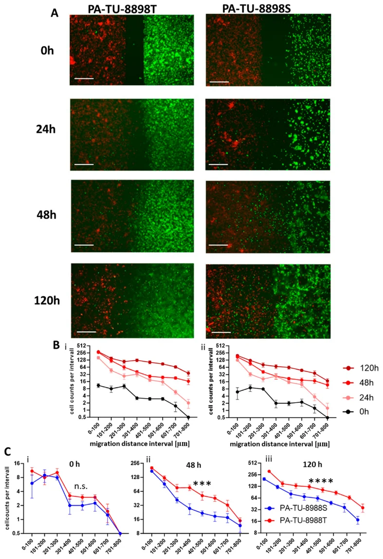
PA-TU-8988T (also designated PaTu 8988t) is a human pancreatic ductal adenocarcinoma cell line established in 1985 from a liver metastasis of a primary pancreatic adenocarcinoma in a 64‑year‑old female patient. It is the poorly differentiated, highly tumorigenic sister line of PA‑TU‑8988S, with both originating from the same metastatic deposit. The line carries driver mutations in KRAS, SMAD4, and TP53, a near‑triploid karyotype (63‑70 chromosomes) and is microsatellite‑stable.
The primary advantage of PA‑TU‑8988T lies in its syngeneic relationship with PA‑TU‑8988S. Despite sharing the same genetic background, the two exhibit sharply contrasting phenotypes: PA‑TU‑8988T forms highly polarized tubular structures in xenografts, expresses abundant mucin, and shows markedly higher transglutaminase activity, indicative of greater differentiation. Most strikingly, PA‑TU‑8988S readily colonizes the lung after intravenous injection, whereas PA‑TU‑8988T fails to metastasize. These lines thus constitute an isogenic model uniquely suited to dissect the molecular underpinnings of differentiation and metastatic competence.
This isogenic pair has been extensively utilized to explore differential glycosylation patterns, galectin‑4‑mediated metastasis restriction, and transcriptional programs controlling autophagy‑lysosome function. PA‑TU‑8988T has also enabled functional validation of stress‑induced apoptosis pathways (e.g., via DDOST or NOX4 manipulation) and drug sensitivity evaluations, advancing studies of pancreatic cancer biology, biomarker discovery, and therapeutic targeting.
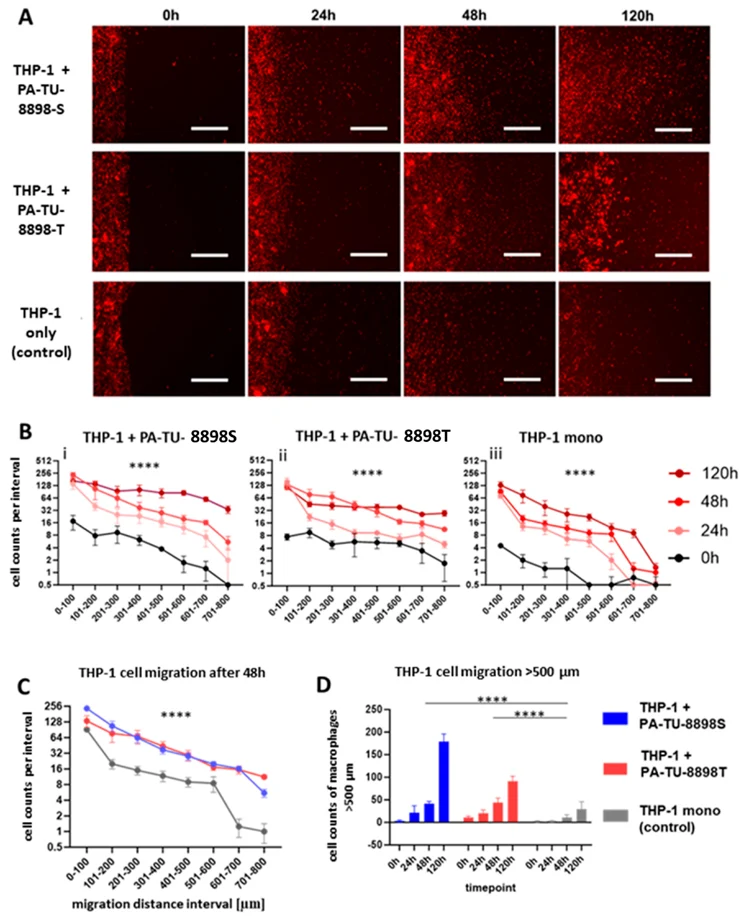
Chamber Gap Assay for Studying Pancreatic Cancer-Induced Macrophage Recruitment and Morphological Alteration
Pancreatic cancer is characterized by an immunosuppressive tumor environment in which macrophages are recruited and reprogrammed to support tumor growth. Studying macrophage migration and polarization is crucial for understanding disease progression and identifying therapeutic targets. However, existing in vitro methods such as the transwell assay provide limited spatial resolution and do not allow visualization of cell movement or morphological changes. Here, Lenz, Maik established and evaluated the Chamber Gap Assay (CGA), a modified two-compartment culture system that enables direct, time-resolved observation and quantification of macrophage migration toward pancreatic cancer cells as well as phenotypic alterations.
Using murine and human macrophage-cancer cell models, we compared the performance of the CGA with the transwell assay. We found that macrophage monocultures displayed substantial spontaneous migration in the transwell system, while cancer cells induced only modest additional macrophage recruitment. In contrast, the CGA demonstrated clear and highly significant directional macrophage movement toward cancer cells, with distinct migration patterns and improved sensitivity for detecting group differences. The method also enabled visualization of cancer cell movement within the same system. Furthermore, CGA offers observations of morphological changes in immune cells under the influence of pancreatic cancer cells.
These findings indicate that the CGA provides a robust and physiologically relevant method for studying tumor-induced immune cell recruitment and associated morphological changes.
Ask a Question
Write your own review
- You May Also Need

Description: Human moderately differentiated adenocarcinoma cell line established from liver metastasis.

Description: Established in 1985 from the primary ductal pancreatic adenocarcinoma (grade II) from a 44-year-old woman; reported as inducing metastasis in nude mice and as secreting proteinases (e.g. urokinase, ...

Description: established from the pancreas of a 81-year-old male patient with pancreatic ductal adenocarcinoma

Description: This cell line is a ductal pancreatic adenocarcinoma derived by differential trypsinisation of explant cultures from a metastatic lesion in the liver of a 26 year old caucasian male with cystic ...


Description: The cells do not express the cystic fibrosis transmembrane conductance regulator (CFTR).
- Adipose Tissue-Derived Stem Cells
- Human Neurons
- Mouse Probe
- Whole Chromosome Painting Probes
- Hepatic Cells
- Renal Cells
- In Vitro ADME Kits
- Tissue Microarray
- Tissue Blocks
- Tissue Sections
- FFPE Cell Pellet
- Probe
- Centromere Probes
- Telomere Probes
- Satellite Enumeration Probes
- Subtelomere Specific Probes
- Bacterial Probes
- ISH/FISH Probes
- Exosome Isolation Kit
- Human Adult Stem Cells
- Mouse Stem Cells
- iPSCs
- Mouse Embryonic Stem Cells
- iPSC Differentiation Kits
- Mesenchymal Stem Cells
- Immortalized Human Cells
- Immortalized Murine Cells
- Cell Immortalization Kit
- Adipose Cells
- Cardiac Cells
- Dermal Cells
- Epidermal Cells
- Peripheral Blood Mononuclear Cells
- Umbilical Cord Cells
- Monkey Primary Cells
- Mouse Primary Cells
- Breast Tumor Cells
- Colorectal Tumor Cells
- Esophageal Tumor Cells
- Lung Tumor Cells
- Leukemia/Lymphoma/Myeloma Cells
- Ovarian Tumor Cells
- Pancreatic Tumor Cells
- Mouse Tumor Cells