
PA-TU-8988S
Cat.No.: CSC-C0326
Species: Homo sapiens (Human)
Source: Liver Metastasis
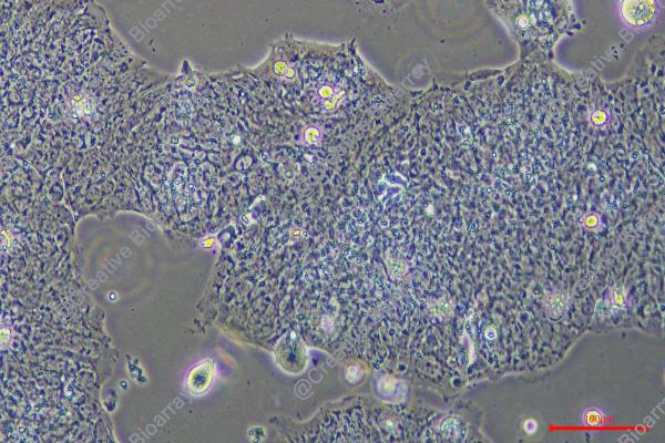
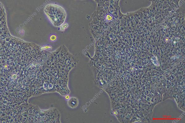
Morphology: adherent epitheloid cells growing in monolayers
Culture Properties: monolayer
- Specification
- Background
- Scientific Data
- Q & A
- Customer Review
Immunology: cytokeratin +, cytokeratin-7 +, cytokeratin-8 +, cytokeratin-17 -, cytokeratin-18 +, cytokeratin-19 +, desmin -, endothel -, EpCAM +, GFAP -, neurofilament -,
PA-TU-8988S (also designated PaTu 8988s) is a well-characterized human pancreatic adenocarcinoma cell line. It was established in 1985 from a liver metastasis of a primary pancreatic adenocarcinoma in a 64-year-old female patient. The cells exhibit adherent epitheloid morphology and grow as monolayers. PA-TU-8988S harbors clinically relevant driver mutations in KRAS, SMAD4, and TP53, mirroring the genetic landscape of human pancreatic ductal adenocarcinoma.
A defining feature of PA-TU-8988S is its origin as one of two sister lines—alongside PA-TU-8988T—derived from the same metastatic lesion. While PA-TU-8988T exhibits a mesenchymal-like, spindle-shaped morphology with high ganglioside levels and low metastatic capacity, PA-TU-8988S displays an epithelial-like phenotype with preserved E-cadherin expression, cell-cell contacts, and polarity. Importantly, PA-TU-8988S is the metastatic counterpart: upon intravenous injection into nude mice, it selectively colonizes the lung, whereas PA-TU-8988T fails to metastasize. This isogenic pair provides an exceptional model for dissecting the molecular determinants of metastasis.
Chamber Gap Assay Enables Sensitive Analysis of Cancer-Induced Macrophage Migration
Pancreatic cancer ranks among the most lethal malignancies. A key contributor to its poor prognosis is the capacity of tumor cells to recruit macrophages and reprogram them into a tumor-promoting rather than tumor-eliminating phenotype. Therefore, elucidating how and why macrophages are recruited to the tumor is essential for designing new therapeutic strategies. However, widely used laboratory methods do not permit direct visualization of cell movement or accurate quantification of migration dynamics. Here, we adapted and evaluated a novel in vitro method, the Chamber Gap Assay (CGA). By culturing cancer cells and macrophages in separate but connected compartments, this system permits real-time visualization of macrophage migration toward cancer cells and the resulting morphological changes.
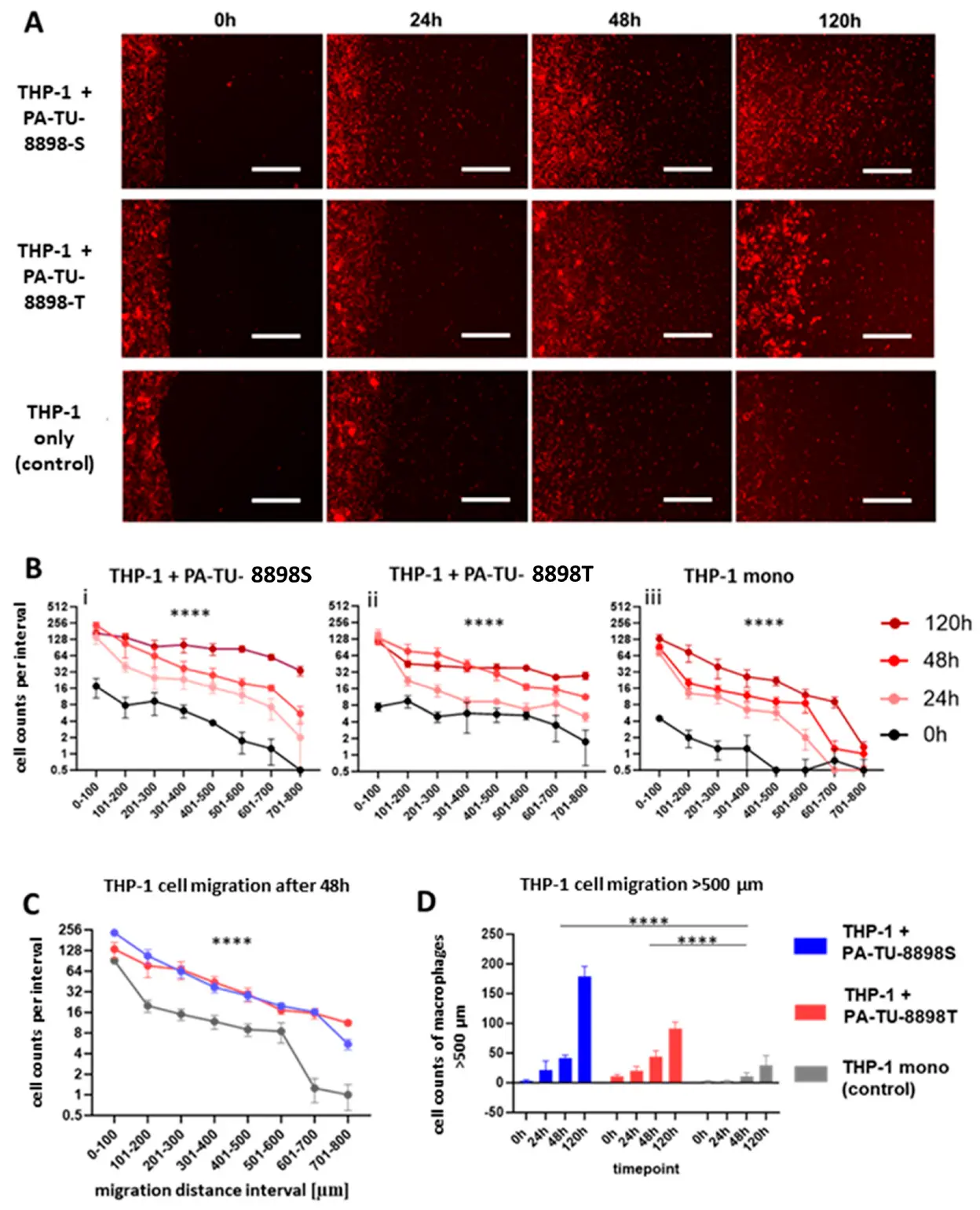
In the model assessing the effects of human pancreatic cancer cell lines PA-TU-8988T and PA-TU-8988S on the migration of THP-1 macrophages, we observed a time-dependent increase in THP-1 macrophage migration (Fig. 1A,B), an increased migration toward pancreatic cancer cells (Fig. 1C), and a gradual rise in the number of migrated THP-1 cells, including cells reaching migration distances beyond 500 µm (Fig. 1D).
Ask a Question
Write your own review
- You May Also Need

Description: Human moderately differentiated adenocarcinoma cell line established from liver metastasis.
Description: Established in 1985 from the primary ductal pancreatic adenocarcinoma (grade II) from a 44-year-old woman; reported as inducing metastasis in nude mice and as secreting proteinases (e.g. urokinase, ...

Description: established from the pancreas of a 81-year-old male patient with pancreatic ductal adenocarcinoma
Description: The MIA PaCa-2 cell line was established from tumor tissue of the pancreas obtained from a 65-year-old Caucasian male in 1975 by A. Yunis, et al. These cells are described to express human colony ...

Description: Established in 2010 from the pancreatic tumor of a 46-year old man with poorly differentiated ductal adenocarcinoma (high grade G3, pT3pN1)
Description: Human tumor cell line secreting parathyroid hormone-related peptide (PTHrP) established from pancreatic tumor.
- Adipose Tissue-Derived Stem Cells
- Human Neurons
- Mouse Probe
- Whole Chromosome Painting Probes
- Hepatic Cells
- Renal Cells
- In Vitro ADME Kits
- Tissue Microarray
- Tissue Blocks
- Tissue Sections
- FFPE Cell Pellet
- Probe
- Centromere Probes
- Telomere Probes
- Satellite Enumeration Probes
- Subtelomere Specific Probes
- Bacterial Probes
- ISH/FISH Probes
- Exosome Isolation Kit
- Human Adult Stem Cells
- Mouse Stem Cells
- iPSCs
- Mouse Embryonic Stem Cells
- iPSC Differentiation Kits
- Mesenchymal Stem Cells
- Immortalized Human Cells
- Immortalized Murine Cells
- Cell Immortalization Kit
- Adipose Cells
- Cardiac Cells
- Dermal Cells
- Epidermal Cells
- Peripheral Blood Mononuclear Cells
- Umbilical Cord Cells
- Monkey Primary Cells
- Mouse Primary Cells
- Breast Tumor Cells
- Colorectal Tumor Cells
- Esophageal Tumor Cells
- Lung Tumor Cells
- Leukemia/Lymphoma/Myeloma Cells
- Ovarian Tumor Cells
- Pancreatic Tumor Cells
- Mouse Tumor Cells