
COV434
Cat.No.: CSC-C9170W
Species: Homo sapiens (Human)
Source: Ovary
Morphology: epithelial
- Specification
- Background
- Scientific Data
- Q & A
- Customer Review
COV434 Cells were isolated from a juvenile granulosa cell tumor (JGCT) of the ovary, which is a type of human ovarian granulosa tumor cell line. COV434 cells have been used extensively as an in vitro system for studying granulosa cell biology, ovarian tumorigenesis, and reproductive endocrinology. COV434 cells grow as adherent cells with an epithelial-like phenotype and express the granulosa cell markers FOXL2, inhibin α, anti-Müllerian hormone (AMH), and aromatase (CYP19A1). They typically grow as monolayers of polygonal cells, though morphology and marker expression may vary depending on conditions used during cell culture. Functionally, they possess features of steroidogenic granulosa cells and secrete factors that help regulate the growth and differentiation of other cells in the ovarian follicle. COV434 cells harbor mutations and activated signaling pathways commonly found in granulosa cell tumors, though some molecular features may differ from models of adult granulosa tumors.
COV434 cells have been used to study processes such as cell proliferation, apoptosis, steroid hormone biosynthesis and signaling, PI3K/AKT signaling, TGF-β/SMAD signaling, cAMP/PKA signaling, and other signaling pathways that regulate granulosa cell function and tumorigenesis. They have been used to study endocrine regulation and ovarian development as well as mechanisms of ovarian tumorigenesis. COV434 cells have also been used in drug screening and evaluation of targeted therapeutics against ovarian granulosa cell tumors.
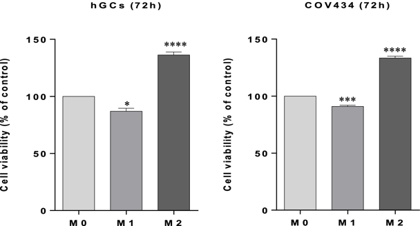
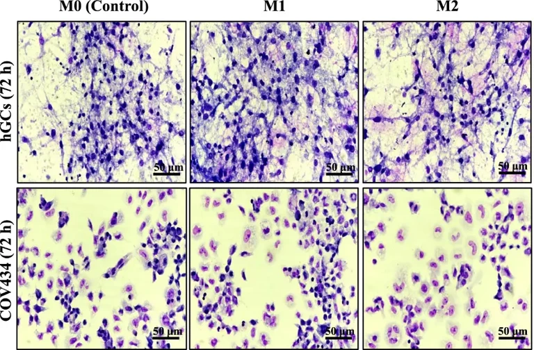
The Role of Macrophages Phenotypes in the Activation of Resolution Pathways within Human Granulosa Cells
Inflammation of the ovaries alters follicular fluid dynamics and leads to poor oocyte quality causing infertility. Macrophages are divided into pro-inflammatory (M1) and anti-inflammatory (M2) phenotypes. These cells secrete inflammatory mediators that could potentially trigger distinct pathways in human granulosa cells (hGCs). However, the impact of these macrophage phenotypes on COX-2 and LOX enzymes expression in hGCs is unknown.
Martins et al. used an in vitro co-culture of hGCs isolated from ART patients with COV434 cell line and stimulated with conditioned media (CM) collected from M0, M1, and M2 macrophages. Real-time PCR and western blot were used to measure COX-2 and LOX enzyme expression levels as markers for oocyte competence. Cell viability assays showed that CM from macrophage M1 and M2 phenotypes altered viability of hGCs and COV434 cells compared to control group M0 after 72 h of interaction (Fig. 1). There were no observed morphological alterations of hGCs or COV434 cells after 72 h of incubation with M1 or M2 CM (Fig. 2). THP-1 macrophage differentiation was confirmed, which allowed for the creation of the interaction model between M1 and M2 macrophage CM and hGCs/COV434 cells. After incubating for 72 h cells were lysed and mRNA and protein were extracted. RT-PCR and western blot analysis was used to analyze COX-2 and 5-, 12-, and 15-LOX expression in hGCs and COV434 cells (Fig. 3).

Ask a Question
Write your own review
- You May Also Need
Description: established from ovary of a 47-year-old female with ovarian endometrioid adenocarcinoma

Description: The A2780 human ovarian cancer cell line was established from tumour tissue from an untreated patient. Cells grow as a monolayer and in suspension in spinner cultures. A2780 is the parent line to the ...
Description: Japanese ovarian serous adenocarcinoma. Said CA125 producing. Cell growth is slow.

Description: Established post-hysterectomy from the tumor tissue of a 65-year-old Japanese woman with malignant ovarian carcinoma (stage: FIGO IIIc; histology: poorly differentiated serous papillary ...

- Adipose Tissue-Derived Stem Cells
- Human Neurons
- Mouse Probe
- Whole Chromosome Painting Probes
- Hepatic Cells
- Renal Cells
- In Vitro ADME Kits
- Tissue Microarray
- Tissue Blocks
- Tissue Sections
- FFPE Cell Pellet
- Probe
- Centromere Probes
- Telomere Probes
- Satellite Enumeration Probes
- Subtelomere Specific Probes
- Bacterial Probes
- ISH/FISH Probes
- Exosome Isolation Kit
- Human Adult Stem Cells
- Mouse Stem Cells
- iPSCs
- Mouse Embryonic Stem Cells
- iPSC Differentiation Kits
- Mesenchymal Stem Cells
- Immortalized Human Cells
- Immortalized Murine Cells
- Cell Immortalization Kit
- Adipose Cells
- Cardiac Cells
- Dermal Cells
- Epidermal Cells
- Peripheral Blood Mononuclear Cells
- Umbilical Cord Cells
- Monkey Primary Cells
- Mouse Primary Cells
- Breast Tumor Cells
- Colorectal Tumor Cells
- Esophageal Tumor Cells
- Lung Tumor Cells
- Leukemia/Lymphoma/Myeloma Cells
- Ovarian Tumor Cells
- Pancreatic Tumor Cells
- Mouse Tumor Cells