
FU-OV-1
Cat.No.: CSC-C0477
Species: Homo sapiens (Human)
Source: Ovary
Morphology: epithelial-like adherent cells growing in monolayers
Culture Properties: monolayer
- Specification
- Background
- Scientific Data
- Q & A
- Customer Review
Immunology: cytokeratin +, cytokeratin-7 -, cytokeratin-8 +, cytokeratin-17 -, cytokeratin-18 +, cytokeratin-19 -, desmin -, endothel -, EpCAM +, GFAP -, neurofilament -,
FU-OV-1 is a human ovarian cancer cell line, which was isolated from the ovarian tumor tissue of a 65-year-old female. She was a patient with malignant ovarian cancer. Her pathological type was FIGO stage IIIc and her histology was poorly differentiated serous papillary adenocarcinoma. The ovary is a part of the female reproductive system, it locates in the pelvic cavity and it is mainly to produce eggs and secrete sex hormones. Ovarian cancer is a tumor that is usually initiated from the epithelial cells on the surface of ovary, and it may also develop in other tissues of ovary. The FU-OV-1 cell line was derived and grown in a serum-free cell culture system. It can be passaged over 96 times. This cell line represents ovarian cancer cells with rapid growth and invasive and metastatic properties. As an ovarian cancer cell line, it is very valuable to study the pathogenesis of ovarian cancer, drug screening and therapeutic strategies.
Identification of Foods that Affect the Anti-Cancer Activity of Pitavastatin in Cells
Statins block mevalonate synthesis, crucial for modifying small GTPase oncogenes. Previously, Jawad et al. showed geranylgeraniol can counteract statins in cell cultures, and pitavastatin reduces ovarian cancer tumors in mice on geranylgeraniol-free diets. Thus, dietary geranylgeraniol might limit statin effectiveness in cancer trials. They identified foods affecting pitavastatin's cytotoxicity in ovarian cancer cells.
Pitavastatin's effect on Ovcar-4 cells was first confirmed (IC50 = 5.2±1.20 µM). Prior findings showed geranylgeraniol and mevalonate, but not farnesol, reduce pitavastatin's cytotoxicity. Further testing revealed that dolichol, coenzyme Q10, and cholesterol did not alter pitavastatin's effect. As expected, geranylgeraniol countered pitavastatin's inhibition. Using Fuov-1 cells, similar patterns appeared with mevalonate pathway metabolites. Next, organic solvent extracts from various foods were tested for geranylgeraniol content. Extracts from oils like corn, sunflower, and grape seed reduced pitavastatin's activity but rape seed oil did not (Fig. 1A). Among fruits and vegetables, beans and cherries partly restored cell growth despite pitavastatin, while lettuce extract significantly suppressed pitavastatin but also stimulated cell growth alone (Fig. 1B). Most carbohydrate-rich food extracts didn't affect pitavastatin, except oats (Fig. 1C). Other foods, including pecan nuts, boiled eggs, and a pasta sauce, suppressed pitavastatin, though some were toxic alone (Fig. 1D). Results were consistent when retested on Fuov-1 cells with selected extracts (Fig. 2).
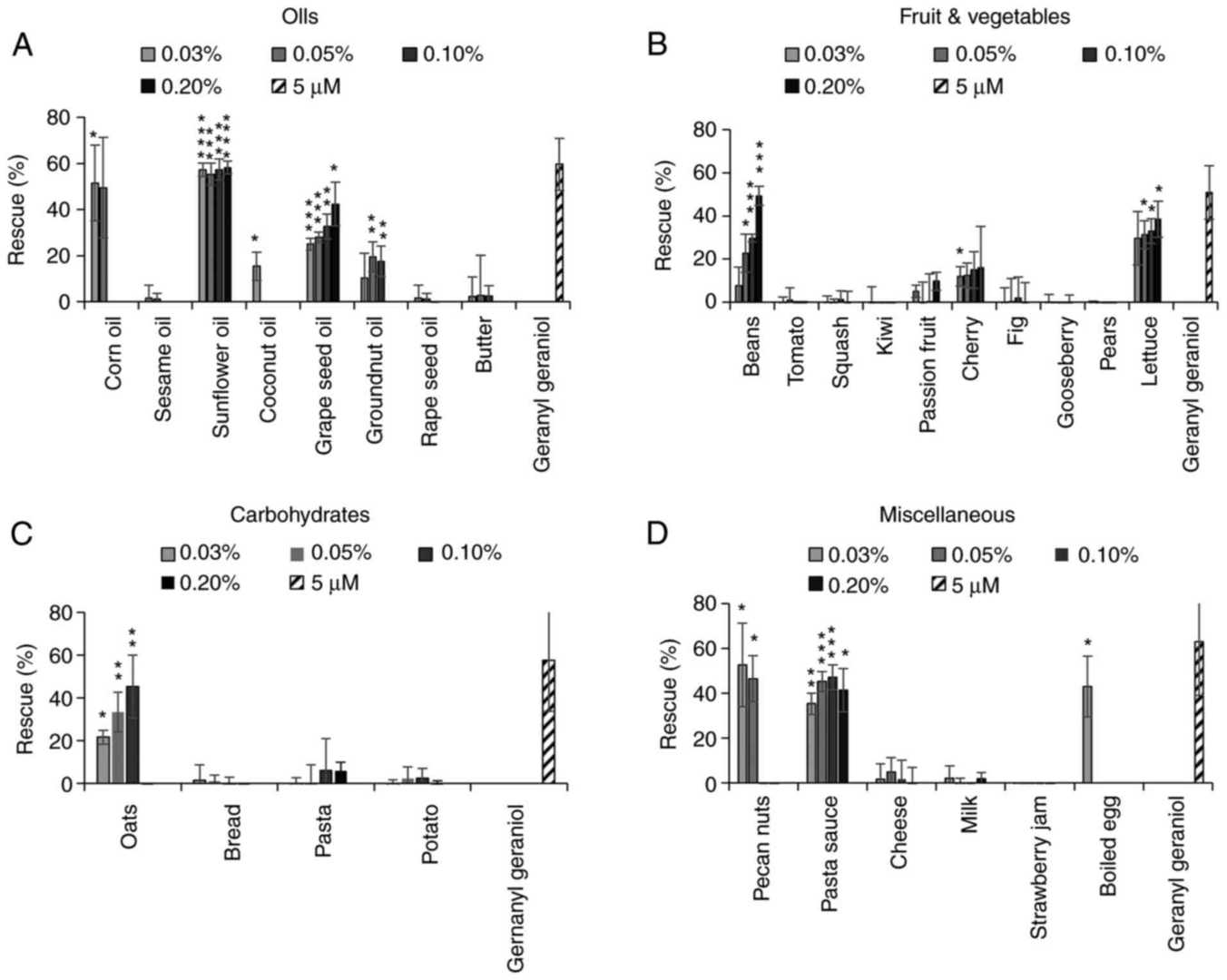 Fig. 1. Effect of food extracts on the activity of pitavastatin in Ovcar-4 cells (Jawad M J, Ibrahim S, et al., 2022).
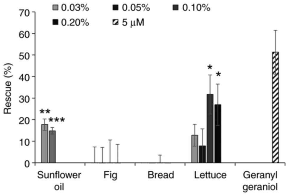 Fig. 2. Effect of food extracts on the activity of pitavastatin in Fuov-1 cells. Effect of extracts from various foodstuffs on the cytotoxic activity of pitavastatin was assessed in Ovcar-4 cells (Jawad M J, Ibrahim S, et al., 2022).
GAB2High Ovarian Cancer Cell Lines are Selectively Sensitive to SHP2 Inhibition
High-grade serous ovarian cancers (HGSOCs) account for the majority of ovarian cancer deaths and pose treatment challenges due to frequent recurrences and platinum resistance. GAB2 overexpression, linked with poor cancer prognoses, facilitates oncogenic signaling in RAS-ERK and PI3K-AKT pathways. To address therapeutic gaps in HGSOCs, Sun et al. employed in vitro and in vivo methods to test dual inhibition of SHP2 and PI3K in GAB2-high ovarian cancer.
They studied if ovarian cancer cell lines with GAB2 amplification/overexpression depend on SHP2 for growth. From CCLE data on 51 cell lines, they identified 9 serous ovarian cancer cell lines with different GAB2 levels. Using PCR and immunoblotting, they found two cell lines (OVCAR3 and JHOS4) with GAB2 amplification and high GAB2 mRNA/protein, two (FUOV1 and IGROV1) with high GAB2 without gene changes, and five (HEYA8, OVCAR5, OV90, OVCAR4, SKOV3) with low GAB2 levels (Fig. 3B and 3C). After SHP099 treatment, the 4 cell lines with high GAB2 showed more sensitivity than the 5 low GAB2 cell lines (Fig. 3D). The were as sensitive or more sensitive than HCC827 lung cancer cells with EGFR mutation to SHP099. SHP099 at 5 μM stopped growth in the 4 high GAB2 lines but not in 2 low GAB2 lines (Fig. 3E and 3F), linked to decreased p-ERK1/2, not p-AKT, levels (Fig. 3G). This suggests SHP2 is necessary for GAB2High ovarian cancer cell growth and ERK1/2 activation.
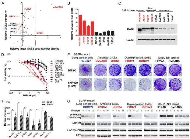 Fig. 3. GAB2High ovarian cancer cell lines are selectively dependent on SHP2 for proliferation and survival (Sun B, Jensen N R, et al., 2019).
Ask a Question
Write your own review
- You May Also Need
Description: established from ovary of a 47-year-old female with ovarian endometrioid adenocarcinoma

Description: The A2780 human ovarian cancer cell line was established from tumour tissue from an untreated patient. Cells grow as a monolayer and in suspension in spinner cultures. A2780 is the parent line to the ...
Description: Japanese ovarian serous adenocarcinoma. Said CA125 producing. Cell growth is slow.


Description: Established from the ascitic fluid of a 56-year-old Caucasian woman with dedifferentiated serous cystadenocarcinoma of the ovary in 1979
- Adipose Tissue-Derived Stem Cells
- Human Neurons
- Mouse Probe
- Whole Chromosome Painting Probes
- Hepatic Cells
- Renal Cells
- In Vitro ADME Kits
- Tissue Microarray
- Tissue Blocks
- Tissue Sections
- FFPE Cell Pellet
- Probe
- Centromere Probes
- Telomere Probes
- Satellite Enumeration Probes
- Subtelomere Specific Probes
- Bacterial Probes
- ISH/FISH Probes
- Exosome Isolation Kit
- Human Adult Stem Cells
- Mouse Stem Cells
- iPSCs
- Mouse Embryonic Stem Cells
- iPSC Differentiation Kits
- Mesenchymal Stem Cells
- Immortalized Human Cells
- Immortalized Murine Cells
- Cell Immortalization Kit
- Adipose Cells
- Cardiac Cells
- Dermal Cells
- Epidermal Cells
- Peripheral Blood Mononuclear Cells
- Umbilical Cord Cells
- Monkey Primary Cells
- Mouse Primary Cells
- Breast Tumor Cells
- Colorectal Tumor Cells
- Esophageal Tumor Cells
- Lung Tumor Cells
- Leukemia/Lymphoma/Myeloma Cells
- Ovarian Tumor Cells
- Pancreatic Tumor Cells
- Mouse Tumor Cells