

Immortalized Rat Brain Microvascular Endothelial Cells
Cat.No.: CSC-I2055Z
Species: rat
Morphology: Polygonal
Culture Properties: Adherent
- Specification
- Background
- Scientific Data
- Q & A
- Customer Review
free from contaminations (bacteria incl. mycoplasma, fungi, HIV, HAV, HBV, HCV, Parvo-B19) and cross-contaminations
Note: Never can cells be kept at -20°C.
Immortalized Rat Brain Microvascular Endothelial Cells are generated from rat cerebral microvascular endothelial tissue and designed to proliferate indefinitely while retaining important endothelial characteristics. These cells are a dependable and repeatable in vitro model for neurovascular research because they retain typical endothelial shape and express distinctive markers linked to the blood-brain barrier (BBB).
Brain microvascular endothelial cells, a vital part of the blood-brain barrier, control the selective exchange of chemicals between the central nervous system and the bloodstream, which is crucial for preserving neural homeostasis. The immortalized form offers improved stability, a longer lifespan, and less batch-to-batch variability as compared to primary cells, which greatly enhances experimental consistency and scalability for long-term studies.
These cells are frequently employed in drug discovery, neuroinflammation research, BBB permeability and transport experiments, and CNS-targeted delivery investigations. In pathological situations such ischemic stroke, neurodegenerative disorders, and brain injury, they are especially useful for examining processes of endothelial barrier function, leukocyte transmigration, and vascular responses. They can also be used in microfluidic organ-on-chip systems for more physiologically relevant research, or co-cultured with astrocytes, pericytes, or neurons to create sophisticated blood-brain barrier or neurovascular unit (NVU) models.
Lasmiditan Induces Mitochondrial Biogenesis in Cerebral Microvascular Endothelial Cells
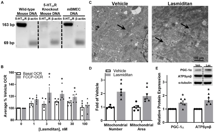
Vascular and mitochondrial dysfunction are hallmarks of central nervous system (CNS) disorders. Given their prior finding that 5-hydroxytryptamine 1F receptor (5-HT1FR) agonism triggers mitochondrial biogenesis (MB), Scholpa et al. investigated the effects of the FDA-approved 5-HT1FR agonist, lasmiditan, in primary mouse brain microvascular endothelial cells (mBMEC). Expression of the target receptor was confirmed prior to experimentation (Fig. 1A).
Treatment with lasmiditan for 48 hours resulted in a concentration-dependent increase in maximal respiratory capacity, measured by FCCP-uncoupled oxygen consumption rate (OCR). A significant ~25% increase was observed at the lowest effective dose of 3 nM (Fig. 1B), which was selected for subsequent experiments. Transmission electron microscopy (Fig. 1C) revealed that lasmiditan-treated cells possessed a greater mitochondrial number and area per field compared to controls (Fig. 1D). Immunoblot analysis corroborated these findings, showing elevated protein levels of the MB regulator PGC-1α and the ETC subunit ATP synthase β (ATPSB) (Fig. 1E), confirming that lasmiditan induces functional MB in mBMEC.
Ask a Question
Write your own review

Description: Immortalized Mouse Primary Artery Fibroblasts-GFP provided by Creative Bioarray have been developed by immortalizing mouse artery fibroblasts with SV40 Large T antigen and transfecting with tGFP. The ...
Description: Immortalized MHC II -/- Mouse Macrophage Cells (C2D) is derived from the knockout mice negative for MHCII molecule. They are stable in culture and survived through crisis in which they spontaneously ...
Description: The bone morphogenetic proteins (BMPs) produced by calvarial cells are crucial in osteoblastic differentiation and bone regeneration, however the limited lifespan of primary cells diminishes their ...
Description: The Immortalized Mouse Atrioventricular Cushion Mesenchymal Cells (tsA58-AVM) were derived from atrioventricular (AV) cushions of H-2Kb -tsA58 embryos at E9.5 and were conditionally immortalized in ...
Description: IDG-SW3 represent a non-homogenous population progressing from early osteoblasts to late osteocytic. These cells express functional SV40 large T antigen that is induced in the presence of IFN γ under ...
Description: The Immortalized Mouse Spleen Dendritic Cell (SRDC) line is functionally and phenotypically similar to dendritic cells; specifically, in its antigen presentation, T cell priming, and dendritic cell ...
- Adipose Tissue-Derived Stem Cells
- Human Neurons
- Mouse Probe
- Whole Chromosome Painting Probes
- Hepatic Cells
- Renal Cells
- In Vitro ADME Kits
- Tissue Microarray
- Tissue Blocks
- Tissue Sections
- FFPE Cell Pellet
- Probe
- Centromere Probes
- Telomere Probes
- Satellite Enumeration Probes
- Subtelomere Specific Probes
- Bacterial Probes
- ISH/FISH Probes
- Exosome Isolation Kit
- Human Adult Stem Cells
- Mouse Stem Cells
- iPSCs
- Mouse Embryonic Stem Cells
- iPSC Differentiation Kits
- Mesenchymal Stem Cells
- Immortalized Human Cells
- Immortalized Murine Cells
- Cell Immortalization Kit
- Adipose Cells
- Cardiac Cells
- Dermal Cells
- Epidermal Cells
- Peripheral Blood Mononuclear Cells
- Umbilical Cord Cells
- Monkey Primary Cells
- Mouse Primary Cells
- Breast Tumor Cells
- Colorectal Tumor Cells
- Esophageal Tumor Cells
- Lung Tumor Cells
- Leukemia/Lymphoma/Myeloma Cells
- Ovarian Tumor Cells
- Pancreatic Tumor Cells
- Mouse Tumor Cells