
Immortalized Mouse Atrioventricular Cushion Mesenchymal Cells (tsA58-AVM)
Cat.No.: CSC-I9355L
Species: Mus musculus
Source: Heart
Morphology: Polygonal
Culture Properties: Adherent
- Specification
- Background
- Scientific Data
- Q & A
- Customer Review
Note: Never can cells be kept at -20 °C.
2) Microarray analysis;
3) RT-PCR analysis.
tsA58-AVM cells are conditionally immortalized mouse Atrioventricular Cushion Mesenchymal Cells (AVM). They were originally isolated from the atrioventricular (AV) cushions of embryonic day 9.5 mouse hearts. Cells derived from these AV cushions are thought to be critical players in heart valve development and are believed to participate in the processes of EndMT as well as valve and septum formation. tsA58 immortalized cells harbor a temperature sensitive allele of SV40 large T antigen (tsA58). Thus, when cells are grown at permissive temperatures (~33 °C), T antigen remains active and allows for prolonged expansion of cells. When cultured at non-permissive temperatures (37-39 °C) inactivation of T antigen allows cells to resemble primary mesenchymal cells more closely.
The tsA58-AVM cells have a spindle-shaped, fibroblast-like morphology and are adherent cells that form monolayers when cultured under normal conditions. The cells are positive for mesenchymal markers including vimentin, α-smooth muscle actin (α-SMA), and N-cadherin. tsA58-AVM cells also maintain functional properties typical of cushion mesenchyme such as migratory capacity, extracellular matrix deposition, and response to developmental cues (TGF-β, BMP, Notch).
Researchers commonly use tsA58-AVM cells to study cardiac cushion development, EndMT, valve morphogenesis and birth defects, and signaling pathways involved in controlling mesenchymal proliferation, differentiation, and extracellular matrix remodeling. Their ability to become immortalized combined with easy growth and maintenance make tsA58-AVM cells an ideal cellular model to study cardiovascular development in vitro.
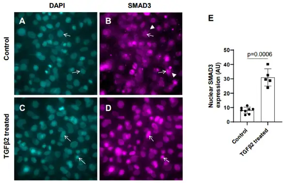
Exogenous TGFβ2 Induced SMAD3 Activation in Cushion Mesenchymal Cells
TGFβ2 signaling together with Hippo signaling play essential roles in cardiac cushion remodeling and loss of TGFβ2 leads to congenital heart defects. Chakrabarti et al. sought to determine if Hippo pathway component YAP1 is mechanistically involved in TGFβ2-mediated ECM remodeling in cushion mesenchymal cells (tsA58-AVM).
In the presence of TGFβ2, SMAD2/3 is unphosphorylated and remains sequestered to the cytoplasm. Phosphorylation of SMAD3 following TGFβ stimulation occurs via receptor-dependent phosphorylation of SMAD3 C-terminal tail, resulting in activation and subsequent translocation to the nucleus to mediate changes in target gene expression and cellular behavior. To determine the effect of TGFβ2 on activation of SMAD3, cushion mesenchymal cells were treated with either 1× PBS or 50 ng/mL TGFβ2 for 12 hours. Immunofluorescence stainings showed SMAD3 primarily localized to the cytoplasm of control cushion mesenchymal cells (Figure 1A, B, arrowheads). Following stimulation with TGFβ2, we observed increased nuclear localization of activated SMAD3 (pSMAD3) (Figure 1C, D, arrows). Quantification of nuclear SMAD3 revealed significant induction upon stimulation with TGFβ2 compared to control (control, n = 8; TGFβ2, n = 5; p = 0.0006) (Figure 1E).
Ask a Question
Write your own review

Description: Immortalized Mouse Primary Artery Fibroblasts-GFP provided by Creative Bioarray have been developed by immortalizing mouse artery fibroblasts with SV40 Large T antigen and transfecting with tGFP. The ...
Description: Immortalized MHC II -/- Mouse Macrophage Cells (C2D) is derived from the knockout mice negative for MHCII molecule. They are stable in culture and survived through crisis in which they spontaneously ...

Description: The bone morphogenetic proteins (BMPs) produced by calvarial cells are crucial in osteoblastic differentiation and bone regeneration, however the limited lifespan of primary cells diminishes their ...
Description: IDG-SW3 represent a non-homogenous population progressing from early osteoblasts to late osteocytic. These cells express functional SV40 large T antigen that is induced in the presence of IFN γ under ...
Description: The Immortalized Mouse Spleen Dendritic Cell (SRDC) line is functionally and phenotypically similar to dendritic cells; specifically, in its antigen presentation, T cell priming, and dendritic cell ...
Description: Immortalized Mouse Primary Dermal Fibroblasts - Adult-GFP provided by Creative Bioarray have been developed by immortalizing mouse dermal fibroblasts with SV40 Large T antigen and transfecting with ...
- Adipose Tissue-Derived Stem Cells
- Human Neurons
- Mouse Probe
- Whole Chromosome Painting Probes
- Hepatic Cells
- Renal Cells
- In Vitro ADME Kits
- Tissue Microarray
- Tissue Blocks
- Tissue Sections
- FFPE Cell Pellet
- Probe
- Centromere Probes
- Telomere Probes
- Satellite Enumeration Probes
- Subtelomere Specific Probes
- Bacterial Probes
- ISH/FISH Probes
- Exosome Isolation Kit
- Human Adult Stem Cells
- Mouse Stem Cells
- iPSCs
- Mouse Embryonic Stem Cells
- iPSC Differentiation Kits
- Mesenchymal Stem Cells
- Immortalized Human Cells
- Immortalized Murine Cells
- Cell Immortalization Kit
- Adipose Cells
- Cardiac Cells
- Dermal Cells
- Epidermal Cells
- Peripheral Blood Mononuclear Cells
- Umbilical Cord Cells
- Monkey Primary Cells
- Mouse Primary Cells
- Breast Tumor Cells
- Colorectal Tumor Cells
- Esophageal Tumor Cells
- Lung Tumor Cells
- Leukemia/Lymphoma/Myeloma Cells
- Ovarian Tumor Cells
- Pancreatic Tumor Cells
- Mouse Tumor Cells