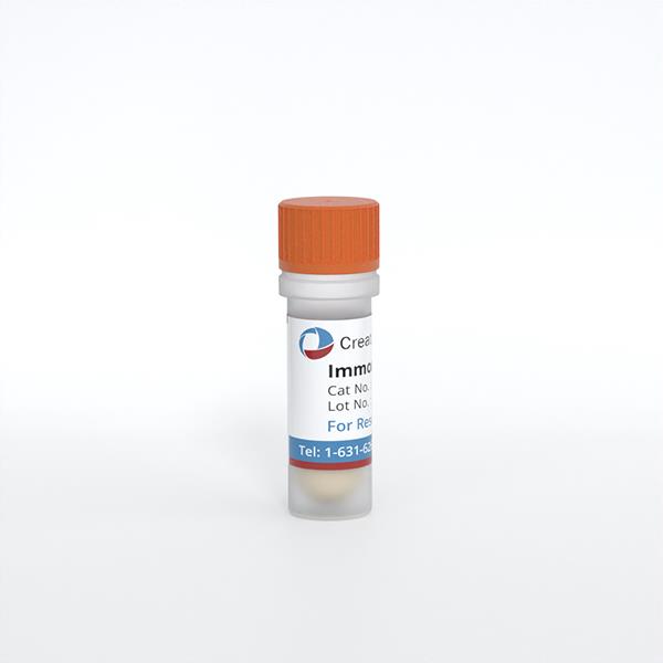
Immortalized (Conditionally) Mouse Osteocytic Cells (IDG-SW3)
Cat.No.: CSC-I9317L
Species: Mus musculus
Source: Long bones
Morphology: Polygonal
Culture Properties: Adherent
- Specification
- Q & A
- Customer Review
Cat.No.
CSC-I9317L
Description
IDG-SW3 represent a non-homogenous population progressing from early osteoblasts to late osteocytic. These cells express functional SV40 large T antigen that is induced in the presence of IFN γ under permissive temperature (33°C) and express GFP under control of the dentin matrix (Dmp1) promoter. Initial culture shows osteoblastic phenotype, however when under mineralizing conditions, the cells start to express early osteocyte markers such as E11/podoplanin, followed by Dmp1 and mature markers SOST and FGF23 by 21-28 days of culture.Similar to osteocytes in vivo, these cells respond to hormonal signals such as PTH and 1,25-dihydroxyvitamin-D3. IDG-SW3 cells can be maintained in both 2D and 3D cultures and are useful for study of osteocytes at various differentiation stages. It should be noted that these cultures may contain a mixture of osteoblasts and osteocytes at different stages of differentiation and for studies requiring enriched osteocytes, the Dmp1-GFP cells should be isolated by flow cytometry (FACS).
Species
Mus musculus
Source
Long bones
Culture Properties
Adherent
Morphology
Polygonal
Immortalization Method
Isolated from long bones of transgenic mice (in which the Dmp1 promoter drives the expression of GFP).
Markers
ALP (osteoblast), E11/gp38, Dmp1 and Phex (osteocyte), SOST/sclerostin and FGF23 (late osteocyte)
Application
For Research Use Only
Storage
Directly and immediately transfer cells from dry ice to liquid nitrogen upon receiving and keep the cells in liquid nitrogen until cell culture needed for experiments.
Note: Never can cells be kept at -20 °C.
Note: Never can cells be kept at -20 °C.
Shipping
Dry Ice.
Recommended Products
CIK-HT003 HT® Lenti-SV40T Immortalization Kit
CIK-HT004 HT® Lenti-SV40 (tsA58 temperature sensitive mutant) Lentivirus Immortalization Kit
CIK-HT004 HT® Lenti-SV40 (tsA58 temperature sensitive mutant) Lentivirus Immortalization Kit
Quality Control
1) Presence and absence of SV40 large T-antigen, under permissive and non-permissive conditions, were confirmed using western blot. 2) Fluorescent imaging was used to measure Dmp1 promoter driven GFP expression in osteogenic conditions.
3) Von Kossa staining was used to show focal nodular mineralization and Alizarin red S staining was used to detect calcium deposition of the differentiated cells.
3) Von Kossa staining was used to show focal nodular mineralization and Alizarin red S staining was used to detect calcium deposition of the differentiated cells.
BioSafety Level
II
Citation Guidance
If you use this products in your scientific publication, it should be cited in the publication as: Creative Bioarray cat no.
If your paper has been published, please click here
to submit the PubMed ID of your paper to get a coupon.
Ask a Question
Write your own review
Related Products
Featured Products
- Adipose Tissue-Derived Stem Cells
- Human Neurons
- Mouse Probe
- Whole Chromosome Painting Probes
- Hepatic Cells
- Renal Cells
- In Vitro ADME Kits
- Tissue Microarray
- Tissue Blocks
- Tissue Sections
- FFPE Cell Pellet
- Probe
- Centromere Probes
- Telomere Probes
- Satellite Enumeration Probes
- Subtelomere Specific Probes
- Bacterial Probes
- ISH/FISH Probes
- Exosome Isolation Kit
- Human Adult Stem Cells
- Mouse Stem Cells
- iPSCs
- Mouse Embryonic Stem Cells
- iPSC Differentiation Kits
- Mesenchymal Stem Cells
- Immortalized Human Cells
- Immortalized Murine Cells
- Cell Immortalization Kit
- Adipose Cells
- Cardiac Cells
- Dermal Cells
- Epidermal Cells
- Peripheral Blood Mononuclear Cells
- Umbilical Cord Cells
- Monkey Primary Cells
- Mouse Primary Cells
- Breast Tumor Cells
- Colorectal Tumor Cells
- Esophageal Tumor Cells
- Lung Tumor Cells
- Leukemia/Lymphoma/Myeloma Cells
- Ovarian Tumor Cells
- Pancreatic Tumor Cells
- Mouse Tumor Cells
Hot Products

