

Immortalized Mouse Intestinal Epithelial Cells-SV40T
Cat.No.: CSC-I2100Z
Morphology: Polygonal
Culture Properties: Adherent
- Specification
- Background
- Scientific Data
- Q & A
- Customer Review
Note: Never can cells be kept at -20°C.
Derived from primary mouse intestinal epithelial cells, Immortalized Mouse Intestinal Epithelial Cells-SV40T are murine cell lines modified genetically with the SV40 large T antigen (SV40T) to enhance their proliferative capacity. Immortalization is achieved through the SV40T protein, which inactivates the key tumor suppressors p53 and retinoblastoma (Rb) proteins. When cultured in the laboratory, these cells exhibit adherent growth and a classic cobblestone shape. They form confluent monolayers with tight cell-to-cell connections, a key characteristic of epithelial cells. These cells also express major intestinal epithelial markers, such as E-cadherin, zonula occludens-1 (ZO-1), villin, and cytokeratins-expressions that verify their epithelial origin and functional barrier abilities. With appropriate culture conditions, the cells can further develop polarized epithelial characteristics and sustain functions associated with intestinal absorption and secretion.
These cells are extensively used in functional research, including investigations into intestinal development, the integrity of the epithelial barrier, immune signaling mechanisms, and cellular reactions to microbial pathogens or inflammatory stimuli. They also play an important role in studies related to drug transport, toxicology, and the modeling of intestinal diseases like inflammatory bowel disease and infection biology. Thanks to their stability, reproducibility, and preserved epithelial phenotype, these cells have become a robust platform for basic gastrointestinal research, translational studies, and preclinical applications.
Calycosin Inhibits NLRP3-Induced Inflammation in IBD
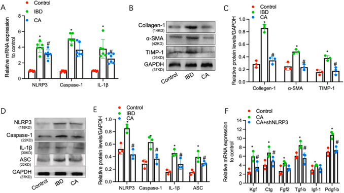
Intestinal interstitial fibrosis is a core event in inflammatory bowel disease (IBD) development. Calycosin possesses various therapeutic bioactivities, but its role in intestinal fibrosis remains unclear. Liao et al. explored calycosin's effects on intestinal interstitial fibrosis and underlying mechanisms using TNBS-induced mouse IBD models, co-culture systems of intestinal epithelial and interstitial cells, and lentivirus-mediated NLRP3 knockdown.
Given that inflammation drives intestinal fibrosis in IBD, they first evaluated NLRP3 expression across control, IBD model, and calycosin-treated (CA) groups. NLRP3 mRNA and its downstream targets caspase-1 and IL-1β were significantly elevated in IBD mice compared to controls, but markedly downregulated by calycosin treatment (Fig. 1A). Similarly, fibrosis markers Collagen-1, TIMP-1, and α-SMA were increased in intestinal epithelial cells from IBD animals and reduced by calycosin (Fig. 1B-C). NLRP3 inflammasome components (NLRP3, caspase-1, ASC) showed comparable patterns (Fig. 1D-E). To elucidate NLRP3's role in calycosin's anti-fibrotic effects, NLRP3 was knocked down in MODE-K intestinal epithelial cells. Calycosin upregulated fibrogenic factors (Kgf, Ctg, Fgf2, Tgf-β, Igf-1, Pdgf-b), but this effect was abolished upon NLRP3 knockdown (Fig. 1F). These results indicate that calycosin modulates fibrogenic factor expression in an NLRP3-dependent manner.
Ask a Question
Write your own review
Description: Immortalized Mouse Primary Artery Fibroblasts-GFP provided by Creative Bioarray have been developed by immortalizing mouse artery fibroblasts with SV40 Large T antigen and transfecting with tGFP. The ...

Description: Immortalized MHC II -/- Mouse Macrophage Cells (C2D) is derived from the knockout mice negative for MHCII molecule. They are stable in culture and survived through crisis in which they spontaneously ...
Description: The bone morphogenetic proteins (BMPs) produced by calvarial cells are crucial in osteoblastic differentiation and bone regeneration, however the limited lifespan of primary cells diminishes their ...
Description: The Immortalized Mouse Atrioventricular Cushion Mesenchymal Cells (tsA58-AVM) were derived from atrioventricular (AV) cushions of H-2Kb -tsA58 embryos at E9.5 and were conditionally immortalized in ...
Description: IDG-SW3 represent a non-homogenous population progressing from early osteoblasts to late osteocytic. These cells express functional SV40 large T antigen that is induced in the presence of IFN γ under ...
Description: The Immortalized Mouse Spleen Dendritic Cell (SRDC) line is functionally and phenotypically similar to dendritic cells; specifically, in its antigen presentation, T cell priming, and dendritic cell ...
- Adipose Tissue-Derived Stem Cells
- Human Neurons
- Mouse Probe
- Whole Chromosome Painting Probes
- Hepatic Cells
- Renal Cells
- In Vitro ADME Kits
- Tissue Microarray
- Tissue Blocks
- Tissue Sections
- FFPE Cell Pellet
- Probe
- Centromere Probes
- Telomere Probes
- Satellite Enumeration Probes
- Subtelomere Specific Probes
- Bacterial Probes
- ISH/FISH Probes
- Exosome Isolation Kit
- Human Adult Stem Cells
- Mouse Stem Cells
- iPSCs
- Mouse Embryonic Stem Cells
- iPSC Differentiation Kits
- Mesenchymal Stem Cells
- Immortalized Human Cells
- Immortalized Murine Cells
- Cell Immortalization Kit
- Adipose Cells
- Cardiac Cells
- Dermal Cells
- Epidermal Cells
- Peripheral Blood Mononuclear Cells
- Umbilical Cord Cells
- Monkey Primary Cells
- Mouse Primary Cells
- Breast Tumor Cells
- Colorectal Tumor Cells
- Esophageal Tumor Cells
- Lung Tumor Cells
- Leukemia/Lymphoma/Myeloma Cells
- Ovarian Tumor Cells
- Pancreatic Tumor Cells
- Mouse Tumor Cells