

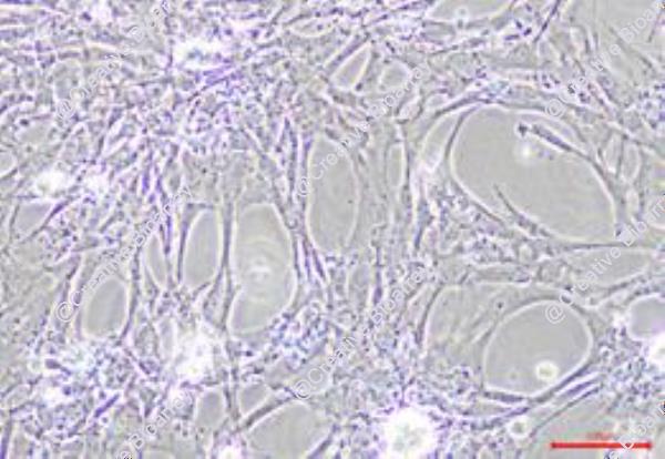
Immortalized Mouse Corneal Epithelial Cells
Cat.No.: CSC-I2070Z
Species: Mouse
Morphology: Polygonal
Culture Properties: Adherent
- Specification
- Background
- Scientific Data
- Q & A
- Customer Review
Note: Never can cells be kept at -20°C.
The corneal epithelium serves as the outermost barrier of the eye, providing a smooth refractive surface while protecting against pathogens, desiccation, and environmental insults. Mouse corneal epithelial cells (MCECs) are widely used to study corneal wound healing, stem cell niche biology, ocular surface homeostasis, and toxicological responses. However, primary MCECs present considerable experimental limitations: they exhibit a restricted proliferative lifespan, undergo rapid senescence in culture, and display phenotypic drift upon passaging. Moreover, isolation from mice is laborious and yields low cell numbers, impeding high-throughput or long-term mechanistic studies.
To circumvent these obstacles, immortalized mouse corneal epithelial cell lines have been established through stable transduction of immortalizing genes, most commonly Simian Virus 40 large T antigen (SV40T) or human telomerase reverse transcriptase (hTERT). This intervention inactivates key cell cycle checkpoints (p53/pRB pathways), enabling continuous proliferation while preserving the fundamental characteristics of their primary counterparts. Immortalized MCECs retain typical cobblestone epithelial morphology, express corneal-specific keratin pairs (K12/K5), and maintain functional intercellular junctions, including tight junctions (ZO-1, occludin) and desmosomes. Importantly, they demonstrate an intact barrier function, appropriate stratification potential under air-lift culture, and responsiveness to exogenous stimuli such as growth factors, cytokines, and chemical irritants.
The use of immortalized mouse corneal epithelial cells offers several distinct advantages: unlimited cell supply, batch-to-batch consistency, ease of genetic manipulation, and suitability for automated screening platforms. Consequently, this cell line provides a robust, reproducible, and ethically advantageous in vitro model for investigating corneal epithelial biology, toxicology, drug permeability, and the molecular mechanisms underlying ocular surface diseases, while eliminating the recurrent need for animal sacrifice.
Cyclosporine A Reprograms Corneal Epithelial TLR2 Signaling to Suppress Dendritic Cell Maturation in Dry Eye Inflammation
This study aimed to elucidate the immunomodulatory mechanism of Cyclosporine A (CsA) in dry eye disease (DED), focusing on its action on corneal epithelial cells (CECs) and the subsequent regulation of dendritic cell (DC) maturation and T-cell activation via the TLR2-MyD88/NF-κB signaling pathway.
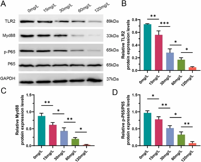
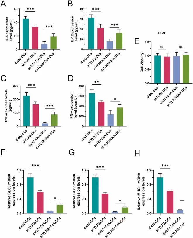
In vitro experiments utilized an immortalized mouse CECs and bone marrow-derived DCs. CECs were treated with a range of CsA concentrations (0-120 mg/L), followed by for TLR2 pathway analysis. TLR2-knockdown CECs were co-cultured with immature DCs to assess DC maturation markers, cytokine secretion, and DC-mediated allogeneic T-cell proliferation.
CsA exposure dose-dependently and potently suppressed TLR2-MyD88/NF-κB signaling in CECs. CsA-treated CECs suppressed DC maturation and reduced the secretion of pro-inflammatory cytokines (IL-6, IL-12, TNF-α, IFN-γ). Consequently, these DCs exhibited a diminished capacity to stimulate T-cell proliferation. Knockdown of TLR2 showed the similar results. However, the inhibitory effect of CsA on DC maturation markers was lost and potentially reversed to a stimulatory phenotype upon TLR2 knockdown in CECs, indicating CEC TLR2 is a primary target for CsA's immunomodulatory effect.
These are mouse-derived corneal epithelial cells that have been genetically modified to be cultured for more than 50 population doublings without showing signs of growth retardation or replicative senescence. They provide a reliable in vitro model for studying corneal epithelial biology and ocular surface research.
Immortalized Mouse Corneal Epithelial Cells (Cat No.: CSC-I2070Z) are ideal for a variety of applications, including ocular surface research, corneal development studies, wound healing investigations, drug testing, and toxicity assessments related to ophthalmic applications.
Unlike primary cells, which have a limited lifespan and batch-to-batch variability, these immortalized cells offer a stable, reproducible model for long-term studies, ensuring consistent experimental results.
Ask a Question
Write your own review

Description: Immortalized Mouse Primary Artery Fibroblasts-GFP provided by Creative Bioarray have been developed by immortalizing mouse artery fibroblasts with SV40 Large T antigen and transfecting with tGFP. The ...
Description: Immortalized MHC II -/- Mouse Macrophage Cells (C2D) is derived from the knockout mice negative for MHCII molecule. They are stable in culture and survived through crisis in which they spontaneously ...

Description: The bone morphogenetic proteins (BMPs) produced by calvarial cells are crucial in osteoblastic differentiation and bone regeneration, however the limited lifespan of primary cells diminishes their ...
Description: The Immortalized Mouse Atrioventricular Cushion Mesenchymal Cells (tsA58-AVM) were derived from atrioventricular (AV) cushions of H-2Kb -tsA58 embryos at E9.5 and were conditionally immortalized in ...
Description: IDG-SW3 represent a non-homogenous population progressing from early osteoblasts to late osteocytic. These cells express functional SV40 large T antigen that is induced in the presence of IFN γ under ...
Description: The Immortalized Mouse Spleen Dendritic Cell (SRDC) line is functionally and phenotypically similar to dendritic cells; specifically, in its antigen presentation, T cell priming, and dendritic cell ...
- Adipose Tissue-Derived Stem Cells
- Human Neurons
- Mouse Probe
- Whole Chromosome Painting Probes
- Hepatic Cells
- Renal Cells
- In Vitro ADME Kits
- Tissue Microarray
- Tissue Blocks
- Tissue Sections
- FFPE Cell Pellet
- Probe
- Centromere Probes
- Telomere Probes
- Satellite Enumeration Probes
- Subtelomere Specific Probes
- Bacterial Probes
- ISH/FISH Probes
- Exosome Isolation Kit
- Human Adult Stem Cells
- Mouse Stem Cells
- iPSCs
- Mouse Embryonic Stem Cells
- iPSC Differentiation Kits
- Mesenchymal Stem Cells
- Immortalized Human Cells
- Immortalized Murine Cells
- Cell Immortalization Kit
- Adipose Cells
- Cardiac Cells
- Dermal Cells
- Epidermal Cells
- Peripheral Blood Mononuclear Cells
- Umbilical Cord Cells
- Monkey Primary Cells
- Mouse Primary Cells
- Breast Tumor Cells
- Colorectal Tumor Cells
- Esophageal Tumor Cells
- Lung Tumor Cells
- Leukemia/Lymphoma/Myeloma Cells
- Ovarian Tumor Cells
- Pancreatic Tumor Cells
- Mouse Tumor Cells