
SH-2
Cat.No.: CSC-C0628
Species: Homo sapiens (Human)
Source: Bone Marrow
Morphology: round cells growing singly and partially in clumps in suspension
Culture Properties: suspension
- Specification
- Background
- Scientific Data
- Q & A
- Customer Review
Immunology: CD3 -, CD4 +, CD13 +, CD14 -, CD15 +, CD19 -, CD33 +, CD34 -, CD56 +
Viruses: PCR: EBV -, HBV -, HCV -, HIV -, HTLV-I/II -, SMRV -
The SH-2 cell line is a human acute myeloid leukemia (AML) cell line established from the bone marrow of a 35-year-old Chinese male patient with AML-M2 (FAB classification) who was refractory to chemotherapy. The cells were initially co-cultured with autologous bone marrow stromal cells and recombinant human interleukin-3 (rhIL-3), and subsequently adapted to grow independently without cytokines or feeder layers.
Morphologically, SH-2 cells are medium-sized round lymphocytes that grow in suspension, either singly or in loose aggregates. They exhibit typical myelomonocytic features with positive peroxidase (POX) staining and express myeloid markers (CD13⁺, CD33⁺, CD117⁺) along with natural killer (NK)-related antigens (CD56⁺, CD16⁺), classifying it as an AML with NK-cell antigen expression. The karyotype is characteristically hypodiploid-45,X,-Y,der(16)t(16;17)(q24;q12),-17,+19-and evolves toward near-tetraploidy with passage. Notably, the line carries hemizygous TP53 deletion (monosomy 17) and a missense mutation (p.Gln192His, c.576G>T) in the remaining TP53allele.
SH-2 cells are routinely cultured in Iscove's Modified Dulbecco's Medium (IMDM) supplemented with serum, at 37°C with 5% CO₂, and are sub-cultured by dilution when reaching approximately 1-2 × 10⁶ cells/mL (doubling time ~80 h). The line is tumorigenic in nude and SCID mice. It is primarily used to investigate AML leukemogenesis, p53-altered chemoresistance mechanisms, and for in vitro drug sensitivity screening in myeloid malignancies.
Relation of NINJ2 and Resistance of RKO Cell to Oxaliplatin
Oxaliplatin is a first-line chemotherapeutic agent for colorectal cancer (CRC), yet intrinsic resistance remains a major obstacle. Ueta et al. hypothesized that Citrobacter freundiimodulates oxaliplatin sensitivity by regulating the expression of the adhesion molecule NINJ2.
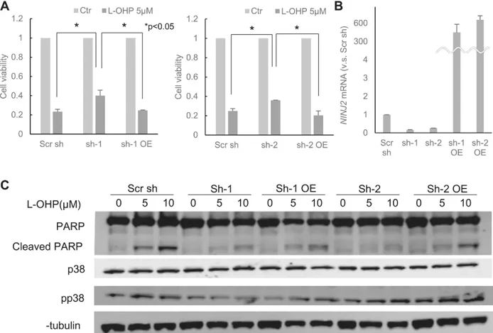
Stable NINJ2-knockdown RKO cell lines (sh-1, sh-2) were established. Knockdown efficacy was confirmed by qPCR (relative expression: Scr sh = 1; sh-1 = 0.16; sh-2 = 0.26; p< 0.01) (Fig. 1B). MTT assays demonstrated that both sh-1 and sh-2 exhibited significantly higher viability upon oxaliplatin treatment compared to the scrambled control (Fig. 1A). Rescue experiments restored NINJ2 expression (Fig. 1B), which significantly reversed resistance, reducing viability compared to the knockdown state (Fig. 1A).
Western blot analysis revealed the mechanistic basis for this resistance (Fig. 1C). In Scr sh control cells, oxaliplatin induced the expression of apoptosis markers (cleaved PARP and phospho-p38) in a dose-dependent manner. This induction was abolished in NINJ2-knockdown cells (sh-1, sh-2). Conversely, restoring NINJ2 expression (sh-1 OE, sh-2 OE) rescued the apoptotic response, reinstating cleaved PARP and pp38 induction. These findings indicate that NINJ2 suppression inhibits oxaliplatin-induced apoptosis, contributing to chemoresistance in CRC.
Ask a Question
Write your own review
- You May Also Need
Description: Established in 2007 from the bone marrow mononuclear cells of an 82-year-old Japanese man with diffuse large B-cell lymphoma in the leukemic phase
Description: Established from the bone marrow of a 28-year-old man who developed the terminal leukemic phase of lymphosarcoma in 1976
Description: This cell line was derived from the bone marrow aspirate of a 59 year old male with erythroleukemia that became acute myelogenous leukaemia.The cells form colonies in soft-agar in the presence of ...
Description: Established from the pleural effusion of a 24-year-old woman with recurrent anaplastic large cell lymphoma (ALCL); cells were described to clonally derive from T-lineage lymphoid cells and to be ...
Description: Established from a 37-year-old man at second (refractory/terminal) relapse of Hodgkin lymphoma (nodular sclerosing -> lymphocyte depleted/stage IIISA -> stage IV) after both combined chemo- and ...
Description: Established from the peripheral blood of a 10-year-old Caucasian boy with acute lymphoblastic leukemia (pre B-ALL) at diagnosis in 1993
- Adipose Tissue-Derived Stem Cells
- Human Neurons
- Mouse Probe
- Whole Chromosome Painting Probes
- Hepatic Cells
- Renal Cells
- In Vitro ADME Kits
- Tissue Microarray
- Tissue Blocks
- Tissue Sections
- FFPE Cell Pellet
- Probe
- Centromere Probes
- Telomere Probes
- Satellite Enumeration Probes
- Subtelomere Specific Probes
- Bacterial Probes
- ISH/FISH Probes
- Exosome Isolation Kit
- Human Adult Stem Cells
- Mouse Stem Cells
- iPSCs
- Mouse Embryonic Stem Cells
- iPSC Differentiation Kits
- Mesenchymal Stem Cells
- Immortalized Human Cells
- Immortalized Murine Cells
- Cell Immortalization Kit
- Adipose Cells
- Cardiac Cells
- Dermal Cells
- Epidermal Cells
- Peripheral Blood Mononuclear Cells
- Umbilical Cord Cells
- Monkey Primary Cells
- Mouse Primary Cells
- Breast Tumor Cells
- Colorectal Tumor Cells
- Esophageal Tumor Cells
- Lung Tumor Cells
- Leukemia/Lymphoma/Myeloma Cells
- Ovarian Tumor Cells
- Pancreatic Tumor Cells
- Mouse Tumor Cells