
MHH-CALL-4
Cat.No.: CSC-C0396
Species: Homo sapiens (Human)
Source: Blood; Peripheral Blood
Morphology: single, round cells growing in suspension, partly in clusters
Culture Properties: suspension
- Specification
- Background
- Scientific Data
- Q & A
- Customer Review
Immunology: CD3 -, CD10 +, CD13 +, CD19 +, CD20 +, CD34 -, CD37 +, CD38 +, CD80 -, CD138 -, HLA-DR +, sm/cyIgG -, sm/cyIgM -, sm/cykappa -, sm/cylambda -
Viruses: ELISA:
MHH-CALL-4 is an acute lymphoblastic leukemia cell line established from the peripheral blood of a 10-year-old boy with pre-B acute lymphoblastic leukemia (pre B-ALL) in 1993. It is a B-cell precursor cell line, and it has many genomic features similar to Ph-like ALL, especially in the aspect of CRLF2 rearrangements and JAK2 mutations. MHH-CALL-4 cells display a small round morphology and predominantly grow in suspension but sometimes form small clusters. MHH-CALL-4 cells have CRLF2 overexpression and the JAK2 I682F mutation, which are responsible for the constitutive activation of the JAK/STAT and PI3K/mTOR signaling pathways in MHH-CALL-4 cells. Studies have reported that JAK2 inhibitors (BBT594) and mTOR inhibitors (AZD2014) potently inhibited JAK/STAT and PI3K/mTOR pathways in MHH-CALL-4 cells with a synergistic effect. TSLP, the ligand of CRLF2, also robustly activated JAK/STAT and PI3K/mTOR pathways to promote cell proliferation and survival in MHH-CALL-4 cells.
Characterization of the MHH-CALL-4 cell line enables several uses in research:
- Leukemia Research: MHH-CALL-4 is used as a model in Leukemia Research to study Ph-like ALL and signaling pathways, especially those associated with CRLF2 rearrangements and JAK2 mutations.
- Drug Screening: The cell line can be used to screen and assess the efficacy of JAK2 and mTOR inhibitors, which have shown strong inhibitory effects.
- Antibody-Mediated Cytotoxicity (ADCC): MHH-CALL-4 cells are used to measure the antibody-mediated cytotoxicity (ADCC) of CD20 antibodies (e.g., rituximab and obinutuzumab) and studies suggest that cell surface sialic acid modification may affect ADCC.
Synergistic Drug Interactions of the Givinostat (ITF2357) in CRLF2-Rearranged Pediatric BCP-ALL Identified by High-Throughput Drug Screening
Pediatric B-cell precursor acute lymphoblastic leukemia (BCP-ALL) with CRLF2 rearrangements is a high-risk subtype, especially in children with Down Syndrome, who often suffer from treatment-related toxicity. Givinostat (ITF2357), a histone deacetylase inhibitor, has shown efficacy against CRLF2-rearranged BCP-ALL. Oikonomou et al. identify synergistic drug combinations with givinostat using high-throughput drug screening to improve treatment outcomes for these patients.
They previously identified givinostat as a promising anti-leukemic agent for CRLF2r BCP-ALL. To evaluate its synergistic potential with other compounds, they used a high-throughput drug screening library of 174 FDA/EMA-approved or preclinical drugs. Using the MHH-CALL-4 cell line, they constructed a per-drug DSS score to assess compound sensitivity in givinostat and DMSO conditions (Fig. 1A). Of the 174 compounds, 54 showed no activity, leaving 120 for further investigation (Fig. 1B). Unsupervised hierarchical clustering distinguished givinostat- and DMSO-treated cells (Fig. 1C), revealing 3 major clusters. One cluster contained drugs whose sensitivity was more modulated by givinostat. A t-test identified 19 compounds with significantly higher sensitivity in the presence of givinostat (Fig. 1D). These compounds belonged to 7 categories: BCL2, FLT3, HSP90, MAPK, PI3K/mTOR, topoisomerase, and p53-MDM2 inhibitors. Setting a DSS threshold of 25, we selected one drug from each category with the highest DSS score in givinostat condition and the largest difference between givinostat and DMSO conditions. The selected drugs were venetoclax, luminespib, trametinib, sapanisertib, etoposide, and idasanutlin (Fig. 1E).
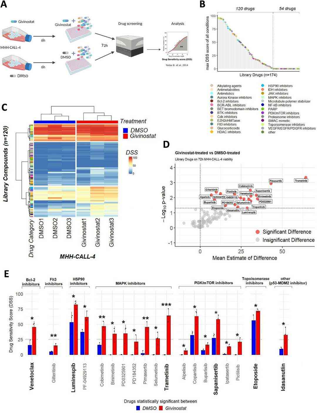 Fig. 1. Givinostat combination high-throughput drug screening on MHH-CALL-4 (Oikonomou A, Watrin T, et al., 2024).
HTS Identifies Synergistic Drug Combinations with Ruxolitinib in Ph-Like ALL
Ph-like ALL is a high-risk leukemia subtype with frequent relapses and poor outcomes, often involving JAK-STAT pathway mutations. CRLF2-rearranged Ph-like ALL show resistance to single-agent JAK inhibitors. Bӧhm et al. identified effective combination treatments and elucidate the mechanisms of synergy for CRLF2-rearranged Ph-like ALL using high-throughput drug screening and molecular analysis.
To identify potential drug combinations with ruxolitinib for Ph-like ALL, they conducted high-throughput screening (HTS) of ruxolitinib with 5057 bioactive compounds against MHH-CALL-4 and MUTZ-5 cell lines, both overexpressing CRLF2 and harboring JAK2, IKZF1, and PAX5 mutations. Despite ruxolitinib inhibiting downstream signaling at 1 µM, both cell lines showed resistance to its cytotoxic effects (IC50 >15 µM for MUTZ-5 and >30 µM for MHH-CALL-4). Thus, they used 1 µM ruxolitinib for HTS. In the primary screen, 460 compounds with <25% survival relative to control were selected for secondary screening. Compounds demonstrating >2-fold enhanced cell kill with ruxolitinib and high single-agent potency were tested in the tertiary screen to estimate IC50 values (Fig. 2A–D). Overall, ~9% of compounds achieved >75% cell death, including kinase inhibitors, glucocorticoids, and anti-neoplastic drugs. Standard-of-care drugs like cyclophosphamide and methotrexate showed limited efficacy, while nucleotide analogs like cytarabine had high single-agent efficacy but only moderate synergy with ruxolitinib.
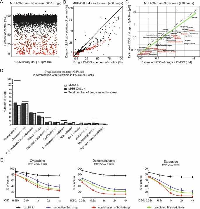 Fig. 2. HTS of the Ph-like ALL cell line MHH-CALL-4 to identify synergistic drug combinations with ruxolitinib (Bӧhm J W, Sia K C S, et al., 2021).
Ask a Question
Write your own review
- You May Also Need

Description: Established in 2007 from the bone marrow mononuclear cells of an 82-year-old Japanese man with diffuse large B-cell lymphoma in the leukemic phase

Description: Established from the bone marrow of a 28-year-old man who developed the terminal leukemic phase of lymphosarcoma in 1976

Description: This cell line was derived from the bone marrow aspirate of a 59 year old male with erythroleukemia that became acute myelogenous leukaemia.The cells form colonies in soft-agar in the presence of ...

Description: Established from the pleural effusion of a 24-year-old woman with recurrent anaplastic large cell lymphoma (ALCL); cells were described to clonally derive from T-lineage lymphoid cells and to be ...

Description: Established from a 37-year-old man at second (refractory/terminal) relapse of Hodgkin lymphoma (nodular sclerosing -> lymphocyte depleted/stage IIISA -> stage IV) after both combined chemo- and ...

Description: Established from the peripheral blood of a 69-year-old woman with B-CLL (chronic lymphocytic leukemia) prior treatment by EBV-transformation in 1988; cell line may represent rather a B-lymphoblastoid ...
- Adipose Tissue-Derived Stem Cells
- Human Neurons
- Mouse Probe
- Whole Chromosome Painting Probes
- Hepatic Cells
- Renal Cells
- In Vitro ADME Kits
- Tissue Microarray
- Tissue Blocks
- Tissue Sections
- FFPE Cell Pellet
- Probe
- Centromere Probes
- Telomere Probes
- Satellite Enumeration Probes
- Subtelomere Specific Probes
- Bacterial Probes
- ISH/FISH Probes
- Exosome Isolation Kit
- Human Adult Stem Cells
- Mouse Stem Cells
- iPSCs
- Mouse Embryonic Stem Cells
- iPSC Differentiation Kits
- Mesenchymal Stem Cells
- Immortalized Human Cells
- Immortalized Murine Cells
- Cell Immortalization Kit
- Adipose Cells
- Cardiac Cells
- Dermal Cells
- Epidermal Cells
- Peripheral Blood Mononuclear Cells
- Umbilical Cord Cells
- Monkey Primary Cells
- Mouse Primary Cells
- Breast Tumor Cells
- Colorectal Tumor Cells
- Esophageal Tumor Cells
- Lung Tumor Cells
- Leukemia/Lymphoma/Myeloma Cells
- Ovarian Tumor Cells
- Pancreatic Tumor Cells
- Mouse Tumor Cells