
CCD-986sk
Cat.No.: CSC-C9353L
Species: Homo sapiens (Human)
Source: Skin
Morphology: fibroblast
Culture Properties: monolayer
- Specification
- Background
- Scientific Data
- Q & A
- Customer Review
vWA: 14,17
FGA: 21,26
Amelogenin: X
TH01: 7,9.3
TPOX: 8,11
CSF1P0: 11,13
D5S818: 11,13
D13S317: 9,12
D7S820: 10,11
Shipping Condition: Room Temperature
CCD-986sk is a human dermal fibroblast cell line established from normal breast skin tissue obtained from a 22-year-old Black female donor during reduction mammoplasty. Its paramount advantage lies in its normal, non-immortalized physiological background-free from oncogenic transformation or disease-associated genetic aberrations-rendering it negative control standard for dermatological and connective tissue studies.
Unlike tumor-derived or virally transformed fibroblast lines, CCD-986sk retains a finite lifespan (senescence ~36 population doublings) and authentic diploid karyology, faithfully recapitulating primary fibroblast behavior. This phenotypic fidelity establishes it as the superior platform for photoaging mechanistics: the line exhibits robust, quantifiable induction of matrix metalloproteinases (MMP-1, MMP-3, MMP-9) and reciprocal suppression of type I procollagen upon UVB/UVA irradiation-responses that mirror human skin chronobiology with high translational validity.
Functionally, CCD-986sk provides a high-throughput screening-compatible matrix for anti-aging compound evaluation. Its validated STR profile (Amelogenin: X; CSF1PO: 11,13; vWA: 14,17) ensure experimental traceability and cross-study reproducibility. The line's exceptional responsiveness to reactive oxygen species (ROS) modulation and extracellular matrix remodeling signals enables precise quantification of pro-collagen synthesis and MMP inhibition, with demonstrated sensitivity to natural product fractions achieving >50% ROS suppression and >50% procollagen mRNA upregulation.
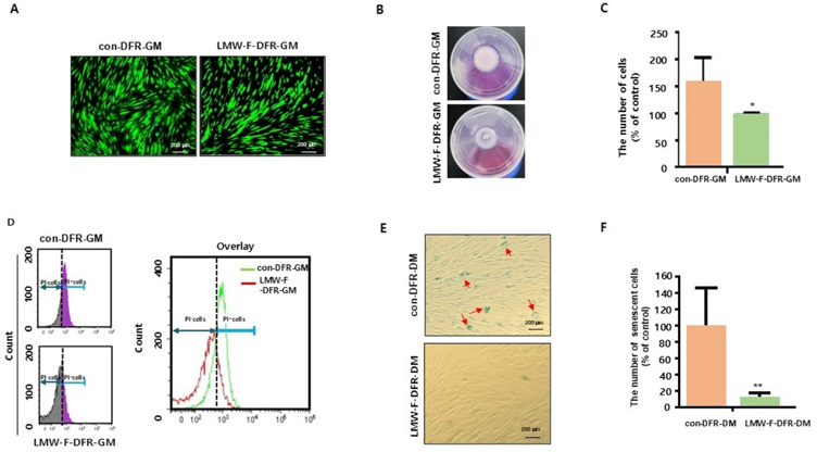
Low-Molecular-Weight Fucoidan Media Enhances Quality and Extents Shelf Life of 3D Human Skin Model
In vitro three-dimensional artificial human skin models (3D-HSMs) have revolutionized research in drug testing, disease modeling, and cosmetic evaluations. However, preserving the shelf-life of 3D-HSM during extended culture remains a challenge.
Previous study uncovered the anti-proliferative effect of fibroblasts exposed to low-molecular-weight fucoidan (LMW-F) at a lower concentration. Based on this study, we formulated two specifically designed media types supplemented with LMW-F components: (1) growth media (DFR-GM) consisting of DMEM (low glucose), Ham's F-12 K, and RPMI 1640 in a precise 2:2:1 ratio, with (LMW-F-DFR-GM) or without LMW-F (con-DFR-GM), and (2) differentiation media (DFR-DM) consisting of the same composition of DFR-GM with the addition of CaCl2, with (LMW-F-DFR-DM) or without LMW-F (con-DFR-DM). To evaluate the effects of LMW-F-DFR-GM on cell proliferation and apoptosis, CCD-986sk cells were incubated with LMW-F-DFR-GM for 7 days. To test the effects of LMW-F-DFR-DM on the aging of fibroblasts, CCD-986sk cells were incubated with LMW-F-DFR-DM for 14 days. Cell proliferation and apoptosis were assessed through live and dead assays, cell counting, and live PI staining with FACS analysis.
The results of the live and dead assays and cell counting revealed that LMW-F-DFR-GM significantly inhibited cell proliferation (approximately 1.5-fold) (Fig.1 A and C). Live PI staining with FACS analysis revealed that the LMW-F-DFR-GM-incubated fibroblasts had a reduced rate of cell death compared to the control group (Fig.1 D). Furthermore, the β-galactosidase analysis revealed that LMW-F-DFR-DM cultivation led to an approximately fivefold reduction in β-gal-positive cells, compared to the control group (Fig.1 E and F). These findings suggest that LMW-F-DFR media may have the potential to enhance the overall dermal health of 3D-FT-HSEMs by decelerating cell proliferation, cell death, and aging in fibroblasts.
Ask a Question
Write your own review

Description: Species: human - male, 60 years old, CaucasianTumorigenecity: Yes, in nude mice; forms malignant melanomaIsoenzyme: PGM3,1;PGM1,1;ES-D,1;AK-1,1;GLO-1,2;G6PD,BHistopathology: malignant melanoma; from ...

Description: Derived from a 14-18 week old human foetus. The cells support growth of CMV and HSV. This cell line has a finite life span.

Description: Species: human male 65 year old; Tissue: skin; Tumor: melanoma; Derived from: a lymph node metastasis

Description: Malignant trichilemmal cyst cells. Strongly laminin positive on cell surface. Serum-free culturable.
Description: Species: human, Caucasian male; Tissue: metastatic cutaneous nodule; Tumor: melanoma

Description: Species: human male; Tissue: subcutaneous metastasis; Tumor: malignant melanoma
- Adipose Tissue-Derived Stem Cells
- Human Neurons
- Mouse Probe
- Whole Chromosome Painting Probes
- Hepatic Cells
- Renal Cells
- In Vitro ADME Kits
- Tissue Microarray
- Tissue Blocks
- Tissue Sections
- FFPE Cell Pellet
- Probe
- Centromere Probes
- Telomere Probes
- Satellite Enumeration Probes
- Subtelomere Specific Probes
- Bacterial Probes
- ISH/FISH Probes
- Exosome Isolation Kit
- Human Adult Stem Cells
- Mouse Stem Cells
- iPSCs
- Mouse Embryonic Stem Cells
- iPSC Differentiation Kits
- Mesenchymal Stem Cells
- Immortalized Human Cells
- Immortalized Murine Cells
- Cell Immortalization Kit
- Adipose Cells
- Cardiac Cells
- Dermal Cells
- Epidermal Cells
- Peripheral Blood Mononuclear Cells
- Umbilical Cord Cells
- Monkey Primary Cells
- Mouse Primary Cells
- Breast Tumor Cells
- Colorectal Tumor Cells
- Esophageal Tumor Cells
- Lung Tumor Cells
- Leukemia/Lymphoma/Myeloma Cells
- Ovarian Tumor Cells
- Pancreatic Tumor Cells
- Mouse Tumor Cells