
HFFF2
Cat.No.: CSC-C2838
Species: Homo sapiens (Human)
Source: Skin; Foreskin
Morphology: Fibroblast
Culture Properties: adherent
- Specification
- Background
- Scientific Data
- Q & A
- Customer Review
CSF1PO: 10,11
D13S317: 11,12
D16S539: 11,13
D5S818: 11
D7S820: 8,12
THO1: 9.3
TPOX: 8,11
vWA: 16,17
Human Fetal Foreskin Fibroblast‑2 (HFFF2) is a normal, diploid fibroblast line established from the foreskin of a Caucasian fetus between 14 and 18 weeks. Morphologically, HFFF2 cells attach to culture vessels as spindle‑shaped, fibroblastic cells with an average diameter of 20-30 µm and a large, round nucleus. The line has a doubling time of 24-30 h in standard culture conditions (DMEM or low‑glucose DMEM with 10 % heat‑inactivated fetal bovine serum, 1 % penicillin/streptomycin, and 5 % CO₂ at 37 °C).
Functionally, HFFF2 cells produce extracellular‑matrix proteins including collagen and fibronectin. The line has been used in studies of wound‑healing and fibroblast‑to‑myofibroblast transition (α‑SMA induction by TGF‑β). HFFF2 is permissive to herpesviruses, particularly CMV and HSV‑1, and has become a standard host cell for viral replication, titration, and antiviral‑drug screening. The line is also broadly used in toxicology assays (MTT/CCK‑8) for testing the cytotoxic effects of nanomaterials, drugs, and photodynamic compounds, as well as in tissue‑engineering research to test cell adhesion and proliferation on biomaterial scaffolds. Co‑culture systems with cancer cells have been used to explore tumor‑stroma interactions, while exposure studies have examined oxidative‑stress responses to UV radiation and 5G‑frequency electromagnetic fields.
Cell Viability of Fe@C/Ole on HFFF2 Cells and hUC-MSCs
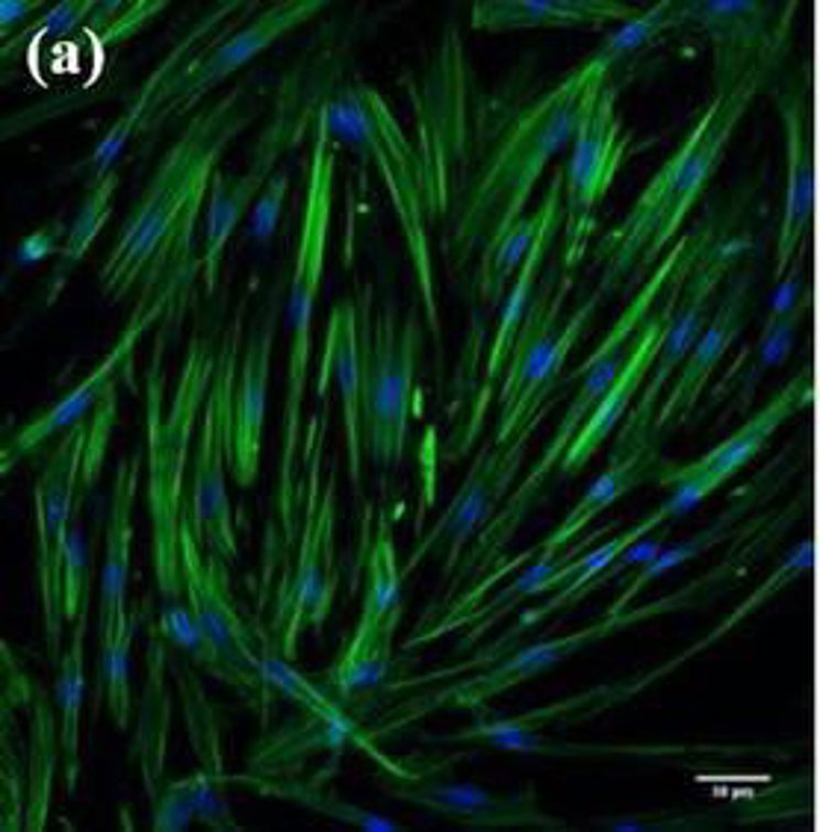
Skin aging, which is affected by intrinsic and extrinsic factors, leads to reduced elastin, collagen, and hydration levels. Safavizadelh et al. utilized modified exosomal content with treated Oleuropein and Fe3O4@C/Oleuropein to modulate gene and microRNA expression on the HFFF2 cells in vitro in order to reduce skin aging.
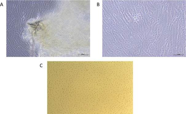
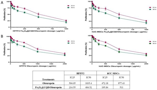
Fe3O4@C/Oleuropein was synthesized via hydrothermal method and confirmed by XRD, FTIR, and SEM. Proliferating cells from human umbilical cord explants exhibited fibroblast-like and spindle-shaped morphologies (Fig. 1A, B). The normal morphology of human dermal fibroblast cells (HFFF2) was observed using phase-contrast microscopy (Fig. 1C). Fe@C/Ole was evaluated for its effects on cell viability (Fig. 2A), with IC50 values of 484.52 μg/mL for HFFF2 cells and 511 μg/mL for hUC-MSCs. Cell treatment was performed at IC25 ≃ 250 μg/mL, which also showed cell growth inhibition. The effects of different Ole concentrations on HFFF2 and hUC-MSC viability were studied (Fig. 2B), with IC50 values of 1035.4 μg/mL for HFFF2 cells and 977.43 μg/mL for hUC-MSCs. Cell treatment was conducted at IC25 ≃ 500 μg/mL as an effective concentration. In summary, Ole exhibited cell inhibition and toxicity at very high concentrations (IC50 ≃ 1000 μg/mL), while Fe@C/Ole showed these effects at lower concentrations (IC25 ≃ 500 μg/mL).
Ask a Question
Write your own review
- You May Also Need

Description: Species: human - male, 60 years old, CaucasianTumorigenecity: Yes, in nude mice; forms malignant melanomaIsoenzyme: PGM3,1;PGM1,1;ES-D,1;AK-1,1;GLO-1,2;G6PD,BHistopathology: malignant melanoma; from ...

Description: Species: human male 65 year old; Tissue: skin; Tumor: melanoma; Derived from: a lymph node metastasis

Description: Malignant trichilemmal cyst cells. Strongly laminin positive on cell surface. Serum-free culturable.
Description: Species: human, Caucasian male; Tissue: metastatic cutaneous nodule; Tumor: melanoma

Description: Species: human male; Tissue: subcutaneous metastasis; Tumor: malignant melanoma

Description: A cell line derived from human skin squamous cell carcinoma having anaplastic epithelioid features.
- Adipose Tissue-Derived Stem Cells
- Human Neurons
- Mouse Probe
- Whole Chromosome Painting Probes
- Hepatic Cells
- Renal Cells
- In Vitro ADME Kits
- Tissue Microarray
- Tissue Blocks
- Tissue Sections
- FFPE Cell Pellet
- Probe
- Centromere Probes
- Telomere Probes
- Satellite Enumeration Probes
- Subtelomere Specific Probes
- Bacterial Probes
- ISH/FISH Probes
- Exosome Isolation Kit
- Human Adult Stem Cells
- Mouse Stem Cells
- iPSCs
- Mouse Embryonic Stem Cells
- iPSC Differentiation Kits
- Mesenchymal Stem Cells
- Immortalized Human Cells
- Immortalized Murine Cells
- Cell Immortalization Kit
- Adipose Cells
- Cardiac Cells
- Dermal Cells
- Epidermal Cells
- Peripheral Blood Mononuclear Cells
- Umbilical Cord Cells
- Monkey Primary Cells
- Mouse Primary Cells
- Breast Tumor Cells
- Colorectal Tumor Cells
- Esophageal Tumor Cells
- Lung Tumor Cells
- Leukemia/Lymphoma/Myeloma Cells
- Ovarian Tumor Cells
- Pancreatic Tumor Cells
- Mouse Tumor Cells