
NBT-T2
Cat.No.: CSC-C6385J
Species: Rattus norvegicus (Rat)
Source: Bladder
Morphology: epithelial-like
Culture Properties: Adherent cells
- Specification
- Background
- Scientific Data
- Q & A
- Customer Review
Store in liquid nitrogen.
NBT-T2 is a non-invasive rat bladder carcinoma cell line derived from the parental NBT-II urothelial carcinoma model. It was established through clonal selection to investigate the molecular mechanisms underlying bladder cancer progression, invasion, and epithelial-mesenchymal transition (EMT). Unlike invasive NBT-II-derived sublines, NBT-T2 retains a stable epithelial phenotype characterized by tight colony formation, strong cell-cell adhesion, and minimal migratory or invasive activity.
NBT-T2 cells are widely used as a comparative model for studying the transition from non-invasive to invasive bladder cancer. The cell line exhibits high expression of epithelial markers and preserved intercellular junctions, making it particularly valuable for investigations of EMT-associated signaling pathways, including TGF-β, EGFR, MAPK, and PI3K/AKT pathways. Researchers frequently employ NBT-T2 alongside invasive NBT-derived variants to identify genetic, epigenetic, and signaling alterations associated with tumor aggressiveness and metastatic potential.
In recent years, NBT-T2 has also been utilized in studies of tumor-microenvironment interactions, immune responses, and therapeutic evaluation in bladder cancer models. Its reproducible epithelial characteristics and well-defined biological behavior make it a reliable platform for anti-cancer drug screening, biomarker discovery, and mechanistic studies of urothelial carcinoma progression. Consequently, NBT-T2 remains an important preclinical model for advancing bladder cancer research and the development of novel therapeutic strategies.
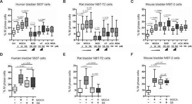
MOCA Induces Mitosis Across Mammalian Bladder Cell Lines Independent of Metabolic Activation
4,4'-Methylenebis(2-chloroaniline) (MOCA), a Group 1 carcinogen, is linked to bladder cancer across species, yet its mechanism remains unclear. While hepatic N-acetyltransferase (NAT) metabolism is traditionally implicated, dogs lack NAT but still develop bladder cancer, suggesting an alternative, metabolism-independent pathway. Kobayashi et al. investigated whether MOCA universally induces mitotic progression in mammalian bladder cells.
They analyzed human (T24, 5637), mouse (MBT-2), and rat (NBT-T2) bladder carcinoma cell lines. MOCA treatment significantly increased the proportion of cells in the M phase compared to control and other aromatic amines, with consistent effects observed even at concentrations as low as 4 µM (Control vs. 4 µM MOCA: p< 0.0001; Fig. 1A-C, F). Co-treatment with rat liver S9 metabolic activation system significantly suppressed MOCA-induced M phase entry (MOCA vs. MOCA+S9: p< 0.0477; Fig. 1D-E), indicating that metabolic processing reduces MOCA's mitotic activity. While S9 alone variably affected baseline mitosis depending on the cell line, the suppressive effect on MOCA was consistent.
These findings demonstrate that MOCA directly induces mitotic progression in mammalian bladder cells across species at environmentally relevant concentrations. The suppression of this effect by metabolic enzymes supports a model wherein parent MOCA, rather than its metabolites, drives chromosomal instability via aberrant mitosis, providing a unifying mechanism for its cross-species carcinogenicity.
Ask a Question
Write your own review
- You May Also Need
Description: Established from the pituitary tumor of an ovarectomized F344 rat treated with estrogen for 3 months; described as showing markedly reduced levels of TGF-β1 and TGF-β type II receptor mRNA; ...
Description: This cell line was derived from a 7-month-old female Wistar-Furth rat. GH3 cells produce growth hormone faster than the GH1 cell line and also produce prolactin. Hydrocortisone stimulates growth ...
Description: medullary thyroid carcinoma, neurotensin and calcitonin-producing, (WAG/RIJ rat)
Description: Ethyl nitrosourea induced tumour of the spinal cord and roots from a Wistar-Furth rat. In cell culture the cells grow to a high density and form a sheet of cells with many processes. TR33B is the ...
- Adipose Tissue-Derived Stem Cells
- Human Neurons
- Mouse Probe
- Whole Chromosome Painting Probes
- Hepatic Cells
- Renal Cells
- In Vitro ADME Kits
- Tissue Microarray
- Tissue Blocks
- Tissue Sections
- FFPE Cell Pellet
- Probe
- Centromere Probes
- Telomere Probes
- Satellite Enumeration Probes
- Subtelomere Specific Probes
- Bacterial Probes
- ISH/FISH Probes
- Exosome Isolation Kit
- Human Adult Stem Cells
- Mouse Stem Cells
- iPSCs
- Mouse Embryonic Stem Cells
- iPSC Differentiation Kits
- Mesenchymal Stem Cells
- Immortalized Human Cells
- Immortalized Murine Cells
- Cell Immortalization Kit
- Adipose Cells
- Cardiac Cells
- Dermal Cells
- Epidermal Cells
- Peripheral Blood Mononuclear Cells
- Umbilical Cord Cells
- Monkey Primary Cells
- Mouse Primary Cells
- Breast Tumor Cells
- Colorectal Tumor Cells
- Esophageal Tumor Cells
- Lung Tumor Cells
- Leukemia/Lymphoma/Myeloma Cells
- Ovarian Tumor Cells
- Pancreatic Tumor Cells
- Mouse Tumor Cells