GS-9L
Cat.No.: CSC-6257W
Species: Rattus norvegicus (Rat)
Source: Brain
Morphology: continuous culture, grown as monolayer, morphology fibroblast-like
Culture Properties: monolayer
- Specification
- Background
- Scientific Data
- Q & A
- Customer Review
Tumor: glioma
GS-9L is a well-known cell line of rat gliosarcoma that came from a brain tumor in Fischer 344 rats that was caused by N-nitrosomethylurea. It grows as a single layer of cells shaped like fibroblasts and stick together. Under normal cell culture conditions, it is usually kept in EMEM medium with fetal bovine serum, L-glutamine, and amino acids that aren't needed. This cell line holds all the characteristics of a malignant cell line, like a fast growth rate, the ability to invade, and the ability to cause tumors in immunocompetent rats of the same type. GS-9L has been extensively utilized as a traditional preclinical model in neuro-oncology research due to its stable biological characteristics and consistent in vivo growth. It makes it easier to study how gliomas grow, invade, angiogenesis, and interact with other cells in the tumor microenvironment. It is also frequently employed to evaluate the efficacy of chemotherapy, radiotherapy, and experimental therapies for malignant brain tumors. GS-9L is a useful tool for studying central nervous system cancers in both living and dead animals because it is based on rodents and cannot fully mimic human glioblastoma.
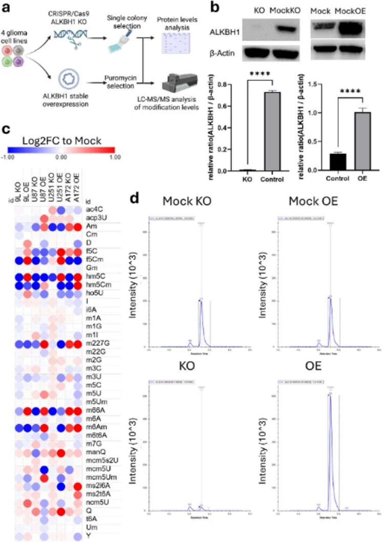
ALKBH1 Slows Proliferation of Glioma Cell In Vitro but Worsens Survival In Vivo
Gliomas rely on translational plasticity for heterogeneity, stemness, and immune evasion; however, the regulatory mechanisms are not well understood. Nakayashiki et al. examined the function of tRNA dioxygenase ALKBH1 in glioma advancement.
Wobble modifications hm5C and f5C extend tRNA-Leu-CAA decoding from Leu-UUG to both Leu-UUG and Leu-UUA codons, potentially impacting translational mechanisms in glioma oncogenesis. To investigate the effects of ALKBH1-mediated wobble oxidation, they created ALKBH1 knockout (KO) and overexpression (OE) models in four glioma cell lines: rodent 9L/GS9L and human U87, U251, and A172 (Fig. 1a). Western blotting showed that the manipulation worked (Fig. 2b). The LC-MS/MS analysis of ALKBH1-associated tRNA modifications (hm5C, hm5Cm, f5C, f5Cm, m1A) demonstrated a consistent decrease in hm5C, hm5Cm, and f5Cm across all knockout lines, with the exception of U251 (Fig. 1c-d). It is important to note that f5C levels did not change after ALKBH1 KO, which suggests that unidentified dioxygenases may be involved. On the other hand, ALKBH1 OE raised hm5C and f5Cm levels in U87, 9L, and U251, but only in U251 did f5C levels go up a lot.
These variations suggest that ALKBH1 activity is dependent on the context, which could be due to differences in the availability of cofactors (like 2-oxoglutarate) or the way tRNA moves in different cells. In glioma cells, their data show that ALKBH1 mainly works as a tRNA wobble dioxygenase, changing m5C to hm5C and f5C at position 34. This is the biochemical basis for codon decoding and translational regulation.
Ask a Question
Write your own review
- You May Also Need
Description: Established from the pituitary tumor of an ovarectomized F344 rat treated with estrogen for 3 months; described as showing markedly reduced levels of TGF-β1 and TGF-β type II receptor mRNA; ...

Description: This cell line was derived from a 7-month-old female Wistar-Furth rat. GH3 cells produce growth hormone faster than the GH1 cell line and also produce prolactin. Hydrocortisone stimulates growth ...

Description: medullary thyroid carcinoma, neurotensin and calcitonin-producing, (WAG/RIJ rat)

Description: Ethyl nitrosourea induced tumour of the spinal cord and roots from a Wistar-Furth rat. In cell culture the cells grow to a high density and form a sheet of cells with many processes. TR33B is the ...
Description: Species: rat - Wistar/FurthTumorigenecity: grow in Wistar/Furth ratsHistopathology: breast cancerNote: Lymph node metastasizing mammary tumor cell line. express Thy-1 and CD8. Metastasizes to all ...
- Adipose Tissue-Derived Stem Cells
- Human Neurons
- Mouse Probe
- Whole Chromosome Painting Probes
- Hepatic Cells
- Renal Cells
- In Vitro ADME Kits
- Tissue Microarray
- Tissue Blocks
- Tissue Sections
- FFPE Cell Pellet
- Probe
- Centromere Probes
- Telomere Probes
- Satellite Enumeration Probes
- Subtelomere Specific Probes
- Bacterial Probes
- ISH/FISH Probes
- Exosome Isolation Kit
- Human Adult Stem Cells
- Mouse Stem Cells
- iPSCs
- Mouse Embryonic Stem Cells
- iPSC Differentiation Kits
- Mesenchymal Stem Cells
- Immortalized Human Cells
- Immortalized Murine Cells
- Cell Immortalization Kit
- Adipose Cells
- Cardiac Cells
- Dermal Cells
- Epidermal Cells
- Peripheral Blood Mononuclear Cells
- Umbilical Cord Cells
- Monkey Primary Cells
- Mouse Primary Cells
- Breast Tumor Cells
- Colorectal Tumor Cells
- Esophageal Tumor Cells
- Lung Tumor Cells
- Leukemia/Lymphoma/Myeloma Cells
- Ovarian Tumor Cells
- Pancreatic Tumor Cells
- Mouse Tumor Cells