
RPMI 6666
Cat.No.: CSC-C9592L
Species: Homo sapiens (Human)
Morphology: lymphoblastoid
Culture Properties: suspension
- Specification
- Background
- Scientific Data
- Q & A
- Customer Review
Species: human - male, 29 years old, Caucasian
Production: immunoglobulin
Histopathology: lymphoma
Note: Hodgkin's disease, EBNA positive; Biological Safety Class II
RPMI 6666 is an established human lymphoblastoid cell line used extensively in both immunology and hematology research. Cells from this line were originally collected from the peripheral blood of a patient suffering from Hodgkin's lymphoma, and are known to grow in suspension and exhibit typical morphology of lymphoblastoid cells. They are generally considered immortalized B-lymphocytes, commonly harboring Epstein-Barr virus as many lines isolated during this time.
In more technical terms, RPMI 6666 cells have been known to express cell surface markers and secrete immunoglobulins, making them ideal for research into B-cell biology, antibody production, and mechanisms of oncogenesis in lymphoma. This cell line is commonly used to research viral-host interactions. The cells have also been used as a substrate for vaccine development and testing of experimental immune-therapeutics. RPMI 6666 can grow consistently in typical media such as RPMI 1640 media supplemented with Fetal Bovine Serum. As such, it acts as a dependable model for malignant lymphoid cells.
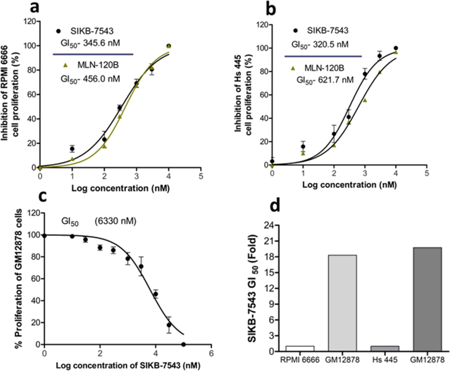
SIKB-7543 Inhibited IKKβ Kinase Activity and Controlled HL Cell Proliferation
HL is aggravated by NF-κB signaling, making IKKβ a target for treatment. Here, Abohassan et al. used high-throughput virtual screening, GROMACS, GMX_MMPBSA, and PLIP to identify leads from the ChemBridge library. They evaluated the in vitro effectiveness of the lead compound, SIKB-7543, using flow cytometry, luminometry, and spectrometry on RPMI 6666 and Hs 445 cells. SIKB-7543 inhibited the proliferation of RPMI 6666 and Hs 445 cell lines with respective GI50 values of 345.6 nM and 320.5 nM (Fig. 1a, b) while the standard MLN 120B had GI50 values of 456 nM and 621.7 nM in RPMI 6666 and Hs 445 cells, respectively (Fig. 1a, b). They next checked the effect of the compound on normal Vero cell proliferation. SIKB-7543 inhibited GM12878 cell proliferation, albeit at a higher GI50 value of 6330 nM (Fig. 1c). The fold concentration of the Vero cell GI50 was 18.31 times higher when compared to the RPMI 6666 cells and 19.75 times higher when compared with Hs 445 cells (Fig. 1d).
Ask a Question
Write your own review
- You May Also Need

Description: Established in 2007 from the bone marrow mononuclear cells of an 82-year-old Japanese man with diffuse large B-cell lymphoma in the leukemic phase

Description: Established from the bone marrow of a 28-year-old man who developed the terminal leukemic phase of lymphosarcoma in 1976

Description: This cell line was derived from the bone marrow aspirate of a 59 year old male with erythroleukemia that became acute myelogenous leukaemia.The cells form colonies in soft-agar in the presence of ...

Description: Established from the pleural effusion of a 24-year-old woman with recurrent anaplastic large cell lymphoma (ALCL); cells were described to clonally derive from T-lineage lymphoid cells and to be ...

Description: Established from a 37-year-old man at second (refractory/terminal) relapse of Hodgkin lymphoma (nodular sclerosing -> lymphocyte depleted/stage IIISA -> stage IV) after both combined chemo- and ...

Description: Established from the peripheral blood of a 10-year-old Caucasian boy with acute lymphoblastic leukemia (pre B-ALL) at diagnosis in 1993
- Adipose Tissue-Derived Stem Cells
- Human Neurons
- Mouse Probe
- Whole Chromosome Painting Probes
- Hepatic Cells
- Renal Cells
- In Vitro ADME Kits
- Tissue Microarray
- Tissue Blocks
- Tissue Sections
- FFPE Cell Pellet
- Probe
- Centromere Probes
- Telomere Probes
- Satellite Enumeration Probes
- Subtelomere Specific Probes
- Bacterial Probes
- ISH/FISH Probes
- Exosome Isolation Kit
- Human Adult Stem Cells
- Mouse Stem Cells
- iPSCs
- Mouse Embryonic Stem Cells
- iPSC Differentiation Kits
- Mesenchymal Stem Cells
- Immortalized Human Cells
- Immortalized Murine Cells
- Cell Immortalization Kit
- Adipose Cells
- Cardiac Cells
- Dermal Cells
- Epidermal Cells
- Peripheral Blood Mononuclear Cells
- Umbilical Cord Cells
- Monkey Primary Cells
- Mouse Primary Cells
- Breast Tumor Cells
- Colorectal Tumor Cells
- Esophageal Tumor Cells
- Lung Tumor Cells
- Leukemia/Lymphoma/Myeloma Cells
- Ovarian Tumor Cells
- Pancreatic Tumor Cells
- Mouse Tumor Cells