
- Specification
- Background
- Scientific Data
- Q & A
- Customer Review
RN46A cells, derived from the embryonic rat brain's raphe nucleus, are an immortalized line known for their abundance of serotonin-releasing neurons. RN46A Cells were immortalized with temperature-sensitive SV40 large T antigen and can be induced to proliferate or differentiate into serotonergic cells depending on the cell culture conditions. RN46A Cells therefore allow for an in vitro study of CNS serotonergic neuron development and function. Differentiated RN46A Cells behave similarly to neuronal cells in that they extend neurite-like processes and express neuronal markers. Furthermore, upon differentiation, RN46A cells display the expression of serotonergic neuron-specific markers, such as tryptophan hydroxylase (TPH), the serotonin transporter (SERT), and several neuronal cytoskeletal proteins.
Due to these characteristics RN46A Cells are used to study serotonin biosynthesis, transport, signaling, neuronal differentiation, neurodevelopment and synaptic regulation. RN46A cells are also a valuable tool for investigating neurotoxicity, neuroinflammation, and how drugs like antidepressants and other psychoactive substances impact serotonin-producing neurons.
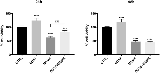
Effects of MDMA and BDNF on Cell Viability by MTT Assay
MDMA ("Ecstasy") causes persistent serotonergic (5-HT) system alterations through unclear molecular mechanisms. Bavato et al. investigated whether BDNF pre-treatment prevents MDMA neurotoxicity and examined MDMA's effects on neurofilament light chain (NfL) in a 5-HT neuronal cell line.
5-HT neurons were differentiated from rat brain raphe nucleus RN46A cells, confirmed by MAP-2 immunocytochemistry (Supplementary Material 4). MDMA IC₅₀ was 1.75 mM at 24 h and 1.15 mM at 48 h; 1.3 mM was selected for subsequent experiments. BDNF (100 ng/ml, 1 h pre-treatment) partially counteracted MDMA-induced cell viability reduction at 24 h (F = 34.68, p < 0.001), but not at 48 h (F = 2.20, p = 0.160) (Fig. 1). Significant differences were found between MDMA and control, and BDNF + MDMA and control, at both time points (p < 0.001).
Ask a Question
Write your own review
Description: Established from the pituitary tumor of an ovarectomized F344 rat treated with estrogen for 3 months; described as showing markedly reduced levels of TGF-β1 and TGF-β type II receptor mRNA; ...

Description: This cell line was derived from a 7-month-old female Wistar-Furth rat. GH3 cells produce growth hormone faster than the GH1 cell line and also produce prolactin. Hydrocortisone stimulates growth ...
Description: medullary thyroid carcinoma, neurotensin and calcitonin-producing, (WAG/RIJ rat)

Description: Ethyl nitrosourea induced tumour of the spinal cord and roots from a Wistar-Furth rat. In cell culture the cells grow to a high density and form a sheet of cells with many processes. TR33B is the ...
- Adipose Tissue-Derived Stem Cells
- Human Neurons
- Mouse Probe
- Whole Chromosome Painting Probes
- Hepatic Cells
- Renal Cells
- In Vitro ADME Kits
- Tissue Microarray
- Tissue Blocks
- Tissue Sections
- FFPE Cell Pellet
- Probe
- Centromere Probes
- Telomere Probes
- Satellite Enumeration Probes
- Subtelomere Specific Probes
- Bacterial Probes
- ISH/FISH Probes
- Exosome Isolation Kit
- Human Adult Stem Cells
- Mouse Stem Cells
- iPSCs
- Mouse Embryonic Stem Cells
- iPSC Differentiation Kits
- Mesenchymal Stem Cells
- Immortalized Human Cells
- Immortalized Murine Cells
- Cell Immortalization Kit
- Adipose Cells
- Cardiac Cells
- Dermal Cells
- Epidermal Cells
- Peripheral Blood Mononuclear Cells
- Umbilical Cord Cells
- Monkey Primary Cells
- Mouse Primary Cells
- Breast Tumor Cells
- Colorectal Tumor Cells
- Esophageal Tumor Cells
- Lung Tumor Cells
- Leukemia/Lymphoma/Myeloma Cells
- Ovarian Tumor Cells
- Pancreatic Tumor Cells
- Mouse Tumor Cells